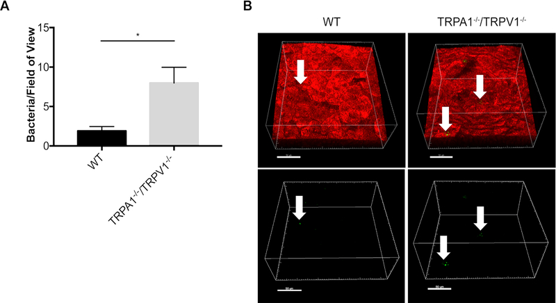
Figure 1.
FISH reveals increased adhesion of environmental bacteria on corneas of TRPA1−/−/TRPV1−/− mice compared to wild-type (WT). A. Quantification of bacteria detected on healthy corneas of WT versus TRPA1−/−/TRPV1−/− by FISH labeling using a universal 16S rRNA gene probe (~ 4-fold difference). Data are expressed as the mean ± SEM number of bacteria per field of view (area of 211μm by 211μm). * P < 0.05 (Student’s t-Test). B. Representative confocal images showing FISH labeling (green, arrows) of environmental bacteria on WT and TRPA1−/−/TRPV1−/− corneas. Upper panels show bacteria (arrows) in the context of the cell membranes of mT/mG mice (red). Lower panels show the same bacteria (green) without cell membrane fluorescence. Scale bar = 50 μm.

