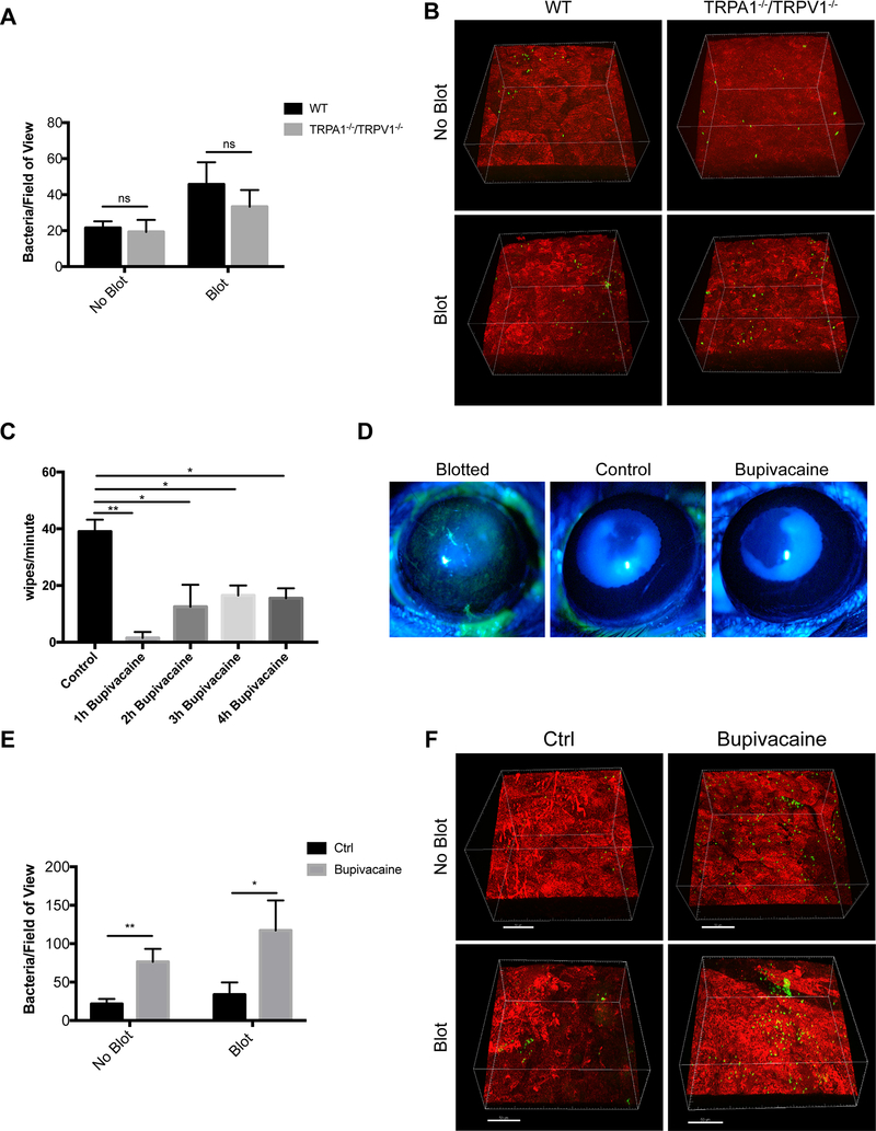
Figure 3.
Evidence for corneal nerve involvement in preventing P. aeruginosa adhesion to the murine cornea. A. Quantification of bacteria adhering to the murine cornea ex vivo after inoculation with ~1×10¹¹ CFU/mL P. aeruginosa on healthy (no blot) and blotted corneas. Ex vivo, equal numbers of P. aeruginosa adhered to TRPA1−/−/TRPV1−/− corneas compared to WT under both healthy and blotted conditions after 4 h. More bacteria adhered to blotted corneas as expected. ns = not significant (Two-way ANOVA). B. Representative images of P. aeruginosa (green) adhering to the cornea (red) in each condition. C. Bupivacaine-treated WT mice showed a significantly diminished response to capsaicin lasting for 4 h compared to mice treated with vehicle control. * P < 0.05, ** P < 0.01 (One-way ANOVA with Tukey’s multiple comparisons test). D. Fluorescein staining of a blotted WT mouse cornea under the slit lamp. Staining was absent in WT mouse corneas treated with either vehicle control or bupivacaine indicating normal epithelial integrity. E. Bupivacaine-treated WT mouse corneas in vivo showed significantly increased adhesion by P. aeruginosa versus vehicle controls under healthy (~ 3.6-fold) and blotted (~ 3.5-fold) conditions. * P < 0.05, ** P < 0.01 (Two-way ANOVA). F. Representative images of P. aeruginosa (green) adhering to the cornea (red) in control and bupivacaine-treated mice. Scale bar = 50 μm.

