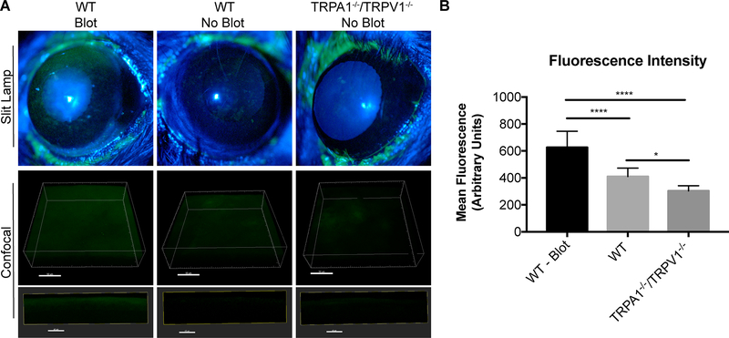
Figure 4.
Intact TRPA1−/−/TRPV1−/− mouse corneas do not stain with fluorescein. A. Fluorescein staining under a slit lamp (upper panels) was evident in blotted corneas of WT mice but absent in healthy corneas (no blot) of WT and TRPA1−/−/TRPV1−/− mice. Confocal images (lower panels) also show that healthy (no blot) corneas of TRPA1−/−/TRPV1−/− and WT mice have little fluorescein staining (and no fluorescein penetration) indicating epithelial integrity was intact compared to blotted WT positive controls. Upper confocal panel images are 3D reconstructions of corneal images (Scale bar = 50 μm) and lower confocal panel images are 10 μM XZ stacks (Scale bar = 40 μm). B. Quantification of mean fluorescence intensity of fluorescein staining in Z-projection of confocal images. Data expressed as the mean ± SEM. **** P < 0.0001, * P < 0.05 (One-way ANOVA with Tukey’s multiple comparisons test).

