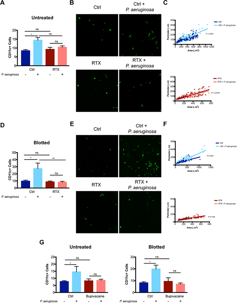
Figure 7.
RTX and bupivacaine attenuate CD11c+ cell infiltration of the murine cornea in response to P. aeruginosa. A. Quantification of CD11c+ cells in healthy (untreated) corneas of control and RTX-treated CD11c-YFP mice at baseline and at 4 h after P. aeruginosa challenge. A significant increase in CD11c+ cells was observed after P. aeruginosa inoculation in control mice that was not observed in RTX-treated mice. * P < 0.05, ns = Not Significant (One-way ANOVA with Tukey’s multiple comparisons test). B. Representative images (maximum intensity projections) of CD11c+ cells in the murine cornea in each condition. C. Morphological analysis of CD11c+ cells using MorpholibJ tools for 3D segmentation in ImageJ and parameters related to z-projections used (perimeter and area) to exclude artifacts due to lower z resolution. Graphs show the distribution of individual cells based on their area and perimeter with a linear regression fit. The upper panel shows a significant difference between curves (P < 0.0001) in control mice with P. aeruginosa inoculation causing increased cell perimeter. The lower panel shows no significant difference between curves (P = 0.078) after P. aeruginosa inoculation in RTX-treated mice. D. Quantification of CD11c+ cells in blotted corneas of control and RTX-treated CD11c-YFP mice at baseline and at 4 h after P. aeruginosa challenge. A significant increase in CD11c+ cells was observed after P. aeruginosa inoculation in controls that was not observed in RTX-treated mice. * P < 0.05, ** P < 0.01, ns = Not Significant (One-way ANOVA with Tukey’s multiple comparisons test). E. Representative images (maximum intensity projections) of CD11c+ cells in blotted corneas in each condition. F. Morphological analysis of blotted corneas showed a significant difference between curves (P < 0.0001) in controls with P. aeruginosa inoculation causing increased cell perimeter, but no difference in curves (P = 0.104) in blotted RTX-treated corneas after P. aeruginosa inoculation. G. Quantification of corneal CD11c+ cells in control and bupivacaine treated mice at baseline and after P. aeruginosa inoculation for 4 h for healthy (untreated) and blotted corneas. For both untreated and blotted corneas, P. aeruginosa inoculation caused a significant increase in CD11c+ cells. Bupivacaine treated corneas showed no response to bacteria. * P < 0.05, ns = Not Significant (One-way ANOVA with Tukey’s multiple comparisons test).

