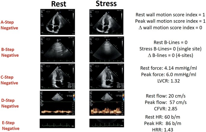
Figure 2.
A normal ABCDE study. Left column: rest images. Right column, stress images. From top to bottom: A step: normal wall motion at rest and during stress; B step: normal A lines in lung ultrasound at rest and during stress; C step: reduced end-systolic volume and increased force during stress; D step: normal increase of pulsed-wave Doppler peak diastolic flow: 56/20 = 2.85; E step: normal heart rate increase on electrocardiogram (86/60 = 1.43). Stress modality: dipyridamole. CFVR, coronary flow velocity reserve; HR, hazard ratio; HRR, heart rate reserve; LVCR, left ventricular contractile reserve.

