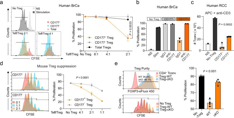
Fig. 7. CD177+ TI Treg cells are highly suppressive to effector T cells.
a Suppressive capacity of CD177+ or CD177− TI Treg cells isolated from breast cancer specimens. CD4 Tconv cells were stimulated by anti-CD3/CD28 co-stimulation. CD4+CD25+CD127low total, CD177+ or CD177− TI Treg cells were purified from 3 individual breast cancer specimens and combined for the suppression assay. Left: Histograms showing CFSE dilution peaks indicating Teff cell proliferation and Right: Percentages of proliferative CD4 Teff cells co-cultured with total, CD177+ or CD177− TI Treg cells at ratios of 2:1, 4:1, 8:1 or no Treg (Combined data from a and b: n = 3 biological replicates for CD177+ TI Treg cells and n = 2 for CD177− TI Treg cells at 2:1 ratio; n = 1 biological replicate for other data point). P value is for comparison between CD177+ or CD177− TI Treg cells at 2:1 ratio Teff/Treg, using two-sided T-test. b Impact of CD177 blockade on the immune suppressive function of CD177+ TI Treg cells, using a monoclonal antibody (MEM166). Similar suppression experiments were performed as in a. CD177+ or CD177− TI Treg cells were purified from fresh human breast cancer specimens (combined from 5 patients) using flow cytometry and co-cultured with effector CD4 T (CD4+CD25−) cells from PBMC for ex vivo suppression assay, with or without the addition of 2 μg/ml isotype control (IgG) or anti-CD177 antibody (MEM166). Percent of proliferating cells were included and the total number of CD4 Tconv cells were enumerated after 4 days of incubation (n = 2 biological replicates for CD177+ Treg group, n = 1 for other data points). c Suppressive capacity of CD177+ or CD177− TI Treg cells isolated from renal cell cancer specimens (RCC). CD4 Tconv cells were stimulated by anti-CD3 and APC (monocyte derived dendritic cells). A total of 7 RCC specimens were combined (n = 1 for NS, non-stimulated or no Treg; n = 4 for CD177+ TI Treg cells; n = 3 biological replicates for CD177− TI Treg cells). The total number of CD4 Tconv cells were counted after 4 days of incubation. d Suppressive capacity of CD177+ or CD177− TI Treg cells isolated from mouse MC38 tumors. Effector CD4 T cells were stimulated by anti-CD3 and APC (derived from T-cell depleted splenocytes of tumor bearing mice) and combined with TI Treg cells at different ratios. Left: Histograms showing the CFSE dilution as an indicator of T cell proliferation and right: Percent proliferation as defined by the histogram (n = 3 biological replicates except n = 6 for no Treg group). Two-way ANOVA was used. e Impact of Treg-specific Cd177-KO on the suppressive capacity of TI Treg cells from MC38 tumors similar as shown in Fig. 5c. Upper left: post-sorting Treg purify was determined by intracellular staining of FoxP3; Lower left: histogram showing CFSE dilution peaks indicating CD4+ Teff cell proliferation; right: summary of percent proliferating cells (n = 4 biological replicates from 14 tumors from cKO group and 20 tumors from WT group, with 4 tumors combined in one biological replicate). All data are presented mean ± s.t.d. c, e one-way ANOVA was used.

