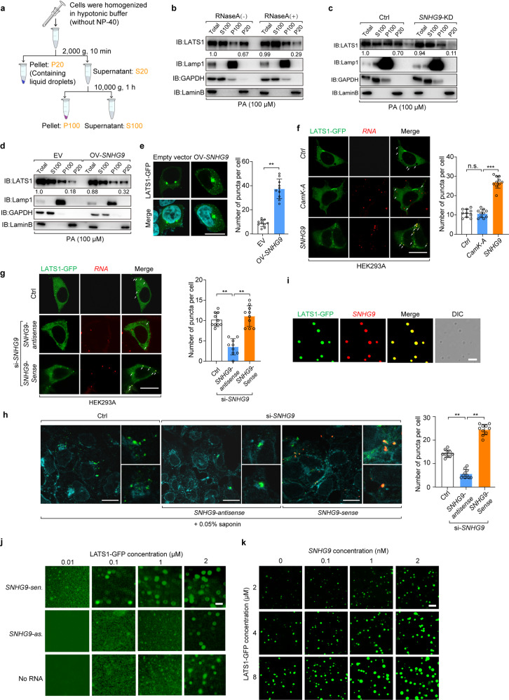
Fig. 5. SNHG9 promotes LATS1 phase separation.
a Schematic diagram of subcellular fractionation procedures. b Subcellular fractionation assay was performed in the presence or absence of RNase A (50 μg/mL) in HEK293A cells. After serum starvation, cells were treated with PA (100 μM) for 1 h. Immunoblot analysis was used to probe the cytosolic fraction (S100), membrane fraction (P100), and nuclear fraction (P20) for the indicated proteins. c Serum-starved wild-type HEK293A cells and SNHG9 KD HEK293A cells, were treated with PA (100 μM) for 1 h. Then subcellular fractionation assay and immunoblot analysis were performed to show the presence of LATS1 in the nuclear fraction (P20). d Serum-starved wild-type HEK293A cells and HEK293A cells overexpressed with SNHG9 were treated with PA (100 μM) for 1 h. Then subcellular fractionation assay and immunoblot analysis were performed to present LATS1 in the nuclear fraction (P20). e HEK293A cells with or without SNHG9 overexpression were transfected with LATS1-GFP (0.5 µg/well, 24-well), and then analyzed by confocal microscopy. Representative pictures were shown (left panel) and the numbers of LATS1-GFP puncta per cell were counted in 10 random fields (right panel). Error bars, SEM of three independent experiments. **P < 0.01, Student’s t-test. Scale bar, 10 μm. f HEK293A cells expressing LATS1-GFP were transfected with exogenous SNHG9 labeled with 546-UTP or CamK-A labeled with 546-UTP, and then analyzed by confocal microscopy. Representative pictures were shown (left panel) and the numbers of LATS1-GFP puncta per cell were counted in 10 random fields. n.s., not significant; ***P < 0.001, Student’s t-test. Scale bar, 10 μm. g HEK293A cells expressing LATS1-GFP were transfected with SNHG9 siRNA or control (Ctrl) siRNA. Cells were transfected with exogenous SNHG9-sen. or SNHG9-as. labeled with 546-UTP, and then analyzed by confocal microscopy. The representative pictures were shown (left panel) and the numbers of LATS1-GFP puncta per cell were counted in 10 random fields (right panel). **P < 0.01, Student’s t-test. Scale bar, 10 μm. h HEK293A cells expressing LATS1-GFP were transfected with SNHG9 siRNA or control RNA. Cells were then transfected with exogenous SNHG9-sen. or SNHG9-as. labeled with 546-UTP, permeabilized with 0.05% saponin and analyzed by confocal microscopy. The representative pictures were shown (left panel) and the numbers of LATS1-GFP puncta per cell were counted in 10 random fields. **P < 0.01, Student’s t-test. Scale bar, 10 μm. i In vitro phase separation assay of LATS1-GFP and in vitro-transcribed SNHG9 labeled with 546-UTP. Scale bar, 2 μm. j In vitro phase separation assay showing droplet formation of LATS1-GFP at different concentrations in the presence of 100 nM SNHG9-sen. (top), 100 nM SNHG9-as. (middle) or no RNA (bottom). Scale bar, 2 μm. k In vitro phase separation assay showing that SNHG9 promotes LATS1 phase separation in a dose-dependent manner. Scale bar, 5 μm.

