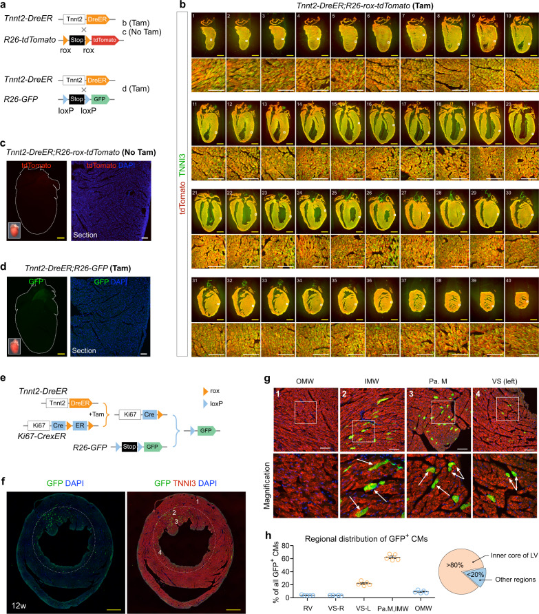
Fig. 4. Tnnt2-DreER triggered ProTracer recording system reveals highly regional cardiomyocyte cell-cycle activity.
a Schematic showing crossing of Tnnt2-DreER with rox or loxP reporters. b Immunostaining for tdTomato and TNNI3 on 1–40 serial heart sections collected from Tnnt2-DreER;R26-rox-tdTomato mice treated with tamoxifen (Tam). Boxed regions are magnified in the lower panels. c Whole-mount and sectional fluorescent images of hearts collected from Tnnt2-DreER;R26-rox-tdTomato mice without tamoxifen treatment (No Tam). d Whole-mount and sectional fluorescent images of hearts collected from Tnnt2-DreER;R26-GFP mice treated with tamoxifen (Tam). e Schematic showing a strategy using Tnnt2-DreER to prime ProTracer system for recording of cardiomyocyte cell-cycle activity. f, g Immunostaining for GFP and TNNI3 on Tnnt2-DreER;Ki67-CrexER;R26-GFP heart sections. Numbered regions (1,2,3,4) are magnified on the (g) panels. Arrows, GFP+ cardiomyocytes. OMW outer myocardial wall, IMW inner myocardial wall, Pa. M papillary muscle, VS ventricular septum. h Quantification of the distribution of GFP+ CMs in different regions of hearts. Data are mean ± s.e.m.; n = 5. Scale bars: yellow, 1 mm; white, 100 µm. Each figure is representative of five individual biological samples.

