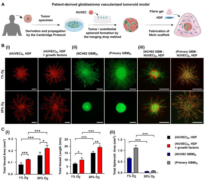
Figure 1.
(A) Schematic representation of the formation of a vascularized tumoroid model from patient-derived primary glioblastoma cells. (B) Fluorescence images showing angiogenic sprouting at 1% and 20% O2 at day 3 after seeding of (i) HUVEC (red) spheroid, supported by single HDF cells with or without additional growth factors; (ii) NCH82 (green) or primary (green) GBM spheroids and (iii) the full vascularized tumoroid model containing NCH82 or primary GBM : HUVEC spheroids with HDF supporting cells. HDF cells are non-fluorescent. The spheroids and single HDF cells are embedded in a 7.5 mg/mL fibrin gel. The scale bars represent 250 m. (C) Graphical representations of (i) the total vessel area and vessel length for HUVEC spheroids surrounded by HDF, with or without additional growth factors and (ii) the total spheroid area for NCH82 or primary GBM spheroids. Data are presented as the mean ± standard deviation; n = 3; *p < 0.05, **p < 0.01, ***p < 0.001. Subscript S denotes cells seeded in the same spheroid.

