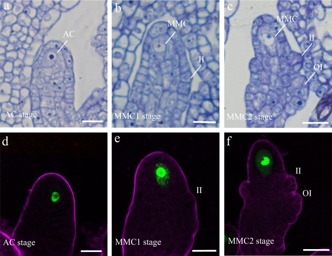
Fig. 1. Ovule primordia structure at the AC, MMC1, and MMC2 stages.
a–c Resin semi-thin sectioning of ovule primordia at the archesporial cell (AC; a) and megaspore mother cell stage 1 (MMC1; b) and MMC2 (c) stage. d–f pKNU:KNU-Venus expression patterns in ovule primordia at the AC (d), MMC1 (e), and MMC2 (f) stages. The magenta signal corresponds to FM4-64 dye outlining the ovule. II inner integument, OI outer integument. Scale bars, 10 μm.

