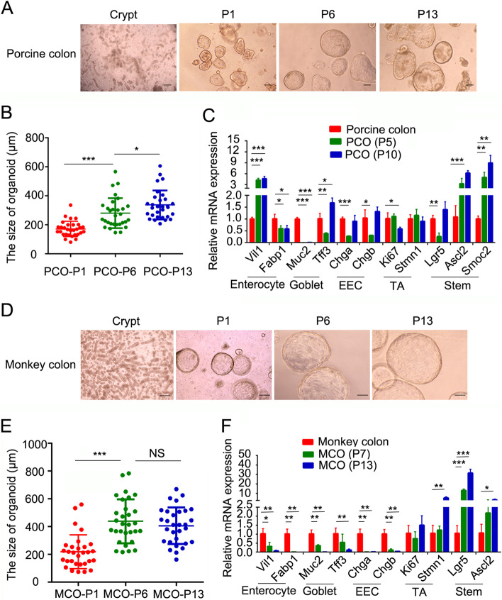
Fig. 1.
Establishment of porcine and monkey colonic organoids. A, D Representative images of organoid growth of porcine colon (A) and monkey colon (D) in expansion medium from different passages. B, E Quantitation of the size of porcine colonic organoids (PCOs) (B) and monkey colonic organoids (MCOs) (E) in different passages. C, F Expression of cell marker genes was examined with q-PCR in primary colon tissue and organoids from different passages in pig (C) and monkey (F). Scale bars, 100 μm. *p < 0.05, ** p < 0.01, ***p < 0.001. Data are displayed as the mean ± SD by one-way ANOVA (B and E) and Student’s t-test (C and F). 30 organoids were calculated for the size in B and E. Three independent experiments were performed in C and F

