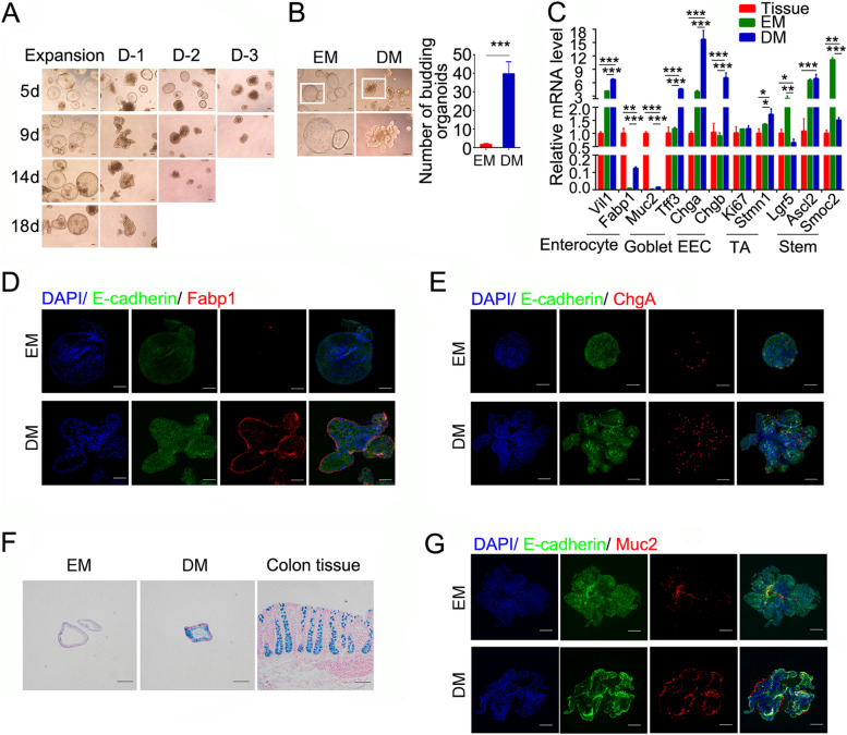
Fig. 2.
Mature lineages of porcine colonic organoids are induced in differentiation medium. A Representative images of porcine colonic organoids grown in different differentiation media. Differentiation medium-1 (D-1): withdrawal of Wnt3a-CM and PGE2 from expansion medium with 2.5 μM CHIR-99021; Differentiation medium-2 (D-2): withdrawal of Wnt3a-CM, PGE2 and CHIR-99021 from expansion medium; Differentiation medium-3 (D-3): withdrawal of Wnt3a-CM, PGE2, CHIR-99021, nicotinamide and SB202190 from expansion medium. B Representative bright-field images and quantitation of the budding numbers of organoids in porcine colonic organoids cultured in expansion or differentiation medium for 7 days. White box depicts higher magnification below. C Expression of cell marker genes in proliferating (EM), differentiated (DM) porcine colonic organoids (passage 15) and tissue. D-G Fabp1 staining (D), ChgA staining (E), Alcian blue staining (F), Muc2 staining (G) in porcine colonic organoids cultured in expansion or differentiation medium for 7 days. Scale bars, 100 μm. *p < 0.05, ** p < 0.01, ***p < 0.001 analyzed by Student’s t-test. Data are shown as mean ± SD (n = 3 independent experiments)

