
Fig. 4.
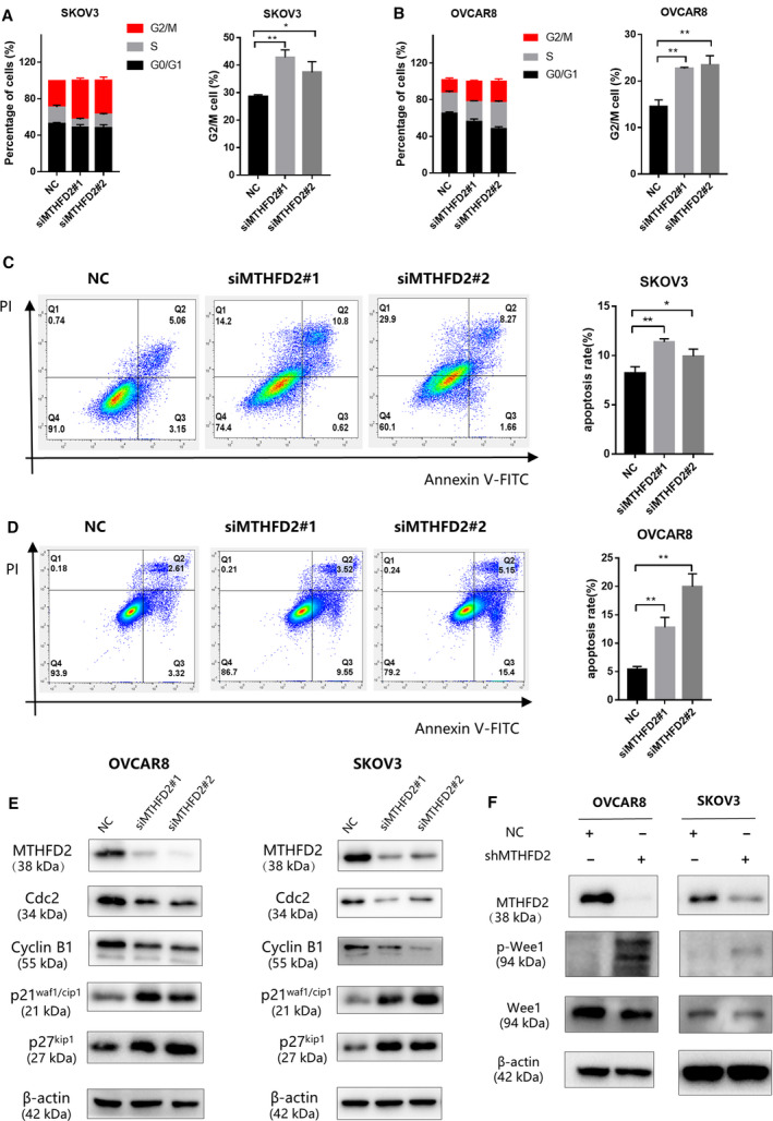
Knockdown of MTHFD2 induces G2/M arrest and cell apoptosis. (A, B) PI staining of SKOV3 and OVCAR8 cells 72 h after transfection with the indicated siRNAs. Flow cytometry analysis shows the cell cycle distribution and G2/M fraction of cells. (C, D) The apoptosis rates of SKOV3 and OVCAR8 cells 72 h after transfection with the indicated siRNAs as evaluated by Annexin V FITC/PI staining. Data of three independent experiments are shown as mean ± standard deviation. (E) The expression of G2/M checkpoint biomarkers, cyclin B1, Cdc2, p21waf1/cip1, p27kip1, proteins were detected by western blot 72 h after transfection into SKOV3 and OVCAR8 cells. The expressions of cyclin B1 and Cdc2 in the MTHFD2 knockdown groups significantly decreased compared with the normal control group. (F) The expression of p‐Wee1 and Wee1 proteins of shMTHFD2 group and shNC group was detected. The expression of p‐Wee1 in the MTHFD2 silencing group is significantly higher than the normal control group. Multiple comparison tests were applied to compare the difference between the groups (*P < 0.05, **P < 0.01).