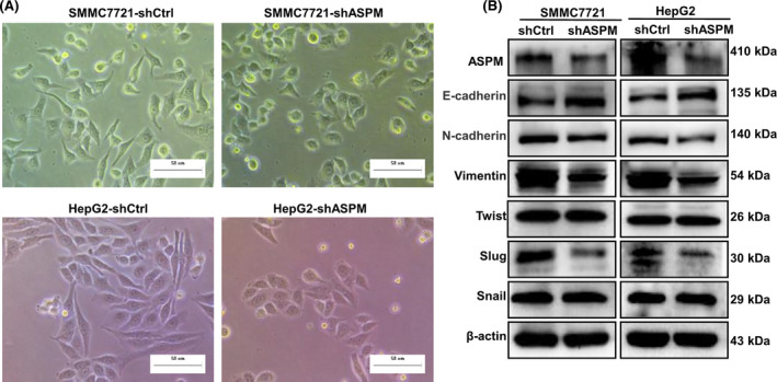
Fig. 4.

ASPM induces EMT in HCC cells. (A) Representative images of the morphological changes of SMMC7721 and HepG2 after silencing of ASPM by shRNA. Scale bars: 50 μm. (B) Western blot analysis of the protein levels of epithelial and mesenchymal markers (E‐cadherin, Vimentin and N‐cadherin), as well as EMT‐related transcriptional factors (Slug, Twist and Snail) in shCtrl or shASPM SMMC7721 and HepG2 cells.
