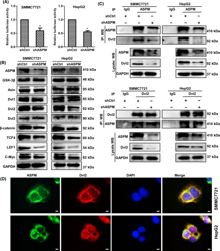
Fig. 5.

ASPM activates Wnt/β‐catenin signaling by interacting with Dvl2 in HCC cells. (A) Luciferase activity of TOP/FOPFlash in control or ASPM‐KD SMMC7721 and HepG2 cells. Data are shown as means ± SD (n = 3 in each assay, Student's t‐test). *P ≤ 0.05 vs. control shRNA. (B) Western blot analysis of the protein expression of Wnt/β‐catenin signaling‐related genes, including GSK‐3β, Axin, Dvl‐2, β‐catenin, TCF4, LEF1 and c‐Myc in control or ASPM‐KD SMMC7721 and HepG2 cells. (C) Reciprocal Co‐IP analysis between ASPM and Dvl2 in control or ASPM‐KD SMMC7721 (top panel) and HepG2 cells (bottom panel). (D) Immunofluorescence analysis of colocalization of ASPM (green) and Dvl‐2 (red) in SMMC7721 and HepG2 cells. Scale bars: 10 μm. IP, immunoprecipitation; WB, western blotting.
