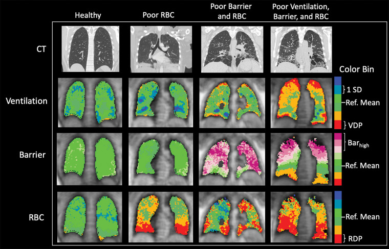
Figure 2:
Representative CT scans, xenon 129 (129Xe) MRI ventilation scans, barrier scans, and red blood cell (RBC) scans in healthy control participant and three participants with nonspecific interstitial pneumonia (NSIP) and range of findings (from left to right: man, age 24 years; woman, age 46 years; woman, age 55 years; and woman, age 57 years). Orange and red indicate reduced signal in ventilation and RBC components, indicating abnormalities. Purple indicates increased signal in barrier component, possibly indicating thickened barrier and/or fibrosis. The first and third scans in participants with NSIP (second and fourth columns in figure) show common pattern of basilar-predominant RBC transfer defects. Note that although these RBC transfer defects are ubiquitous features in participants with NSIP, some participants with NSIP have preserved, normal-looking barrier uptake and/or ventilation. Regional correspondence is visible between CT and 129Xe MRI abnormalities. Notably, regions of high barrier measured using 129Xe MRI are also associated with normal CT findings, suggesting early-stage microstructural disease activity not yet visible on CT scan. Barhigh = high barrier percentage, Ref. = reference, RDP = red blood cell defect percentage, SD = standard deviation, VDP = ventilation defect percentage.

