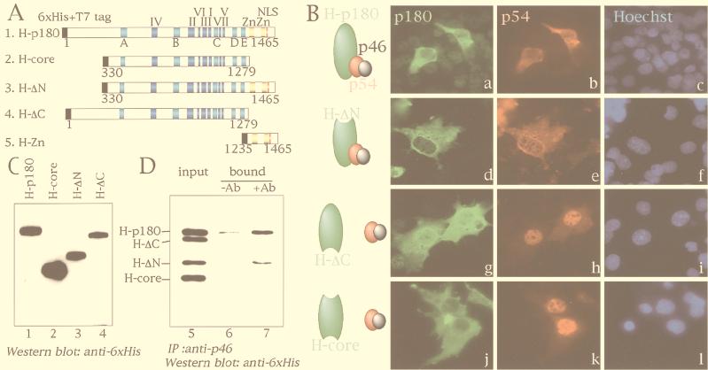
FIG. 3.
Identification of the p180 domain required for the interaction with p54-p46. (A) Schematic representation of p180 mutant constructs. H-p180, H-ΔN, H-ΔC, and H-core indicate full-length p180 with six-His tag and T7 tag at the amino terminus, amino-terminally deleted p180 with six-His tag and T7 tag at the amino terminus, carboxyl-terminally deleted p180 with six-His tag and T7 tag at the amino terminus, and both amino-terminally and carboxyl-terminally deleted p180 with six-His tag and T7 tag at the amino terminus, respectively. H-Zn, which contains only carboxyl-terminal putative zinc finger regions, is also shown for the experiments shown in Fig. 4. The seven highly conserved regions of class B DNA polymerases are indicated by blue boxes with roman numerals (I to VII) (32, 43). The five conserved regions in eukaryotic DNA polymerase α are indicated by light blue boxes with letters (A to E) (22). Putative zinc finger motifs and a putative NLS (23) are depicted by yellow boxes and a red line, respectively. Six-His and T7 tags are shown by solid boxes. Numbers indicate amino acid positions of p180. (B) Subcellular distribution of ectopically expressed p180 mutants and p54-p46. H-p180 (a to c), H-ΔN (d to f), H-ΔC (g to i), and H-core (j to l) were cotransfected with pI-pri into COS-1 cells, and the expressed proteins were detected simultaneously by indirect immunofluorescence analysis with anti-p54 polyclonal antibody and Texas red-conjugated anti-rabbit IgG antibody or anti-six-His monoclonal antibody and FITC-conjugated anti-mouse IgG antibody. Nuclear staining with Hoechst is shown in the rightmost panels. (C) Western blot analysis of transiently expressed p180 mutants. Extracts (10 μg of protein) were subjected to SDS-PAGE followed by Western blot analysis with anti-six-His monoclonal antibody. Lane numbers correspond to p180 mutant constructs shown in panel A. (D) Coimmunoprecipitation assay. Extracts (50 μg of protein) containing p54-p46 were mixed with a total of 250 μg of the extracts described for panel C (75 μg of the extracts containing H-p180, 75 μg of the extracts containing H-ΔN, 75 μg of the extracts containing H-ΔC, and 25 μg of the extracts containing H-core), incubated on ice for 2 h, and immunoprecipitated with (lane 7) or without (lane 6) anti-p46 antibody and protein G-Sepharose. One-third of the precipitates were subjected to Western blot analysis with anti-six-His monoclonal antibody. Lane 5 contains 5 μg of the input proteins. Ab, antibody; IP, immunoprecipitation.

