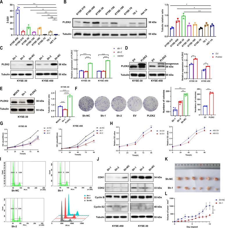
Fig. 2. Silence of PLEK2 inhibited proliferation of ESCC cells in vivo and vitro.
A Expression of PLEK2 in ESCC cell lines detected by qPCR; B Western blot analysis of level of PLEK2 in several ESCC cell lines; C Silence of PLEK2 expression in KYSE-30 and KYSE-450. Western Blot was used to confirm the knockdown efficiency; D Overexpression of PLEK2 expression in KYSE-30 and KYSE-450 Western Blot was used to confirm the overexpression efficiency; E Reverse effect of PLEK2 overexpression plasmid on PLEK2 stably knockdown KYSE-30 and KYSE-450; F The colony generated by KYSE-30 with or without PLEK2 stably knockdown, as well as KYSE-30 with or without PLEK2 stably overexpressed. G Proliferation ability of stably knockdown PLEK2 in KYSE-30 and KYSE-450 detected by CCK-8 assay; H Proliferation ability of stably overexpression PLEK2 in KYSE-30 and KYSE-450 detected by CCK-8 assay; I Flow cytometry diagrams of cell cycle of both KYSE-30 with or without PLEK2 stably knockdown. J Western blot analysis for the expression of CDK1, CDK2, Cyclin B, and Cyclin E2, which related to cell cycle. Tubulin was used as loading control; K Representative xenograft tumours derived from mice subcutaneously injected with KYSE-30 cells with or without PLEK2 stably knockdown. (n = 6 in each group). L The growth curve of tumours in two groups (n = 6 in each group). *P < 0.01; ***P < 0.01; ***P < 0.001.

