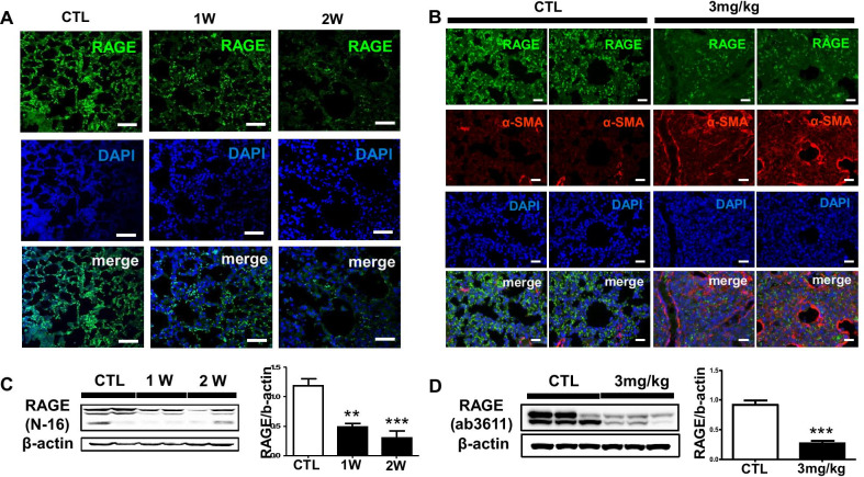
Fig. 3.
RAGE expression is down-regulated in BLM mouse model of pulmonary fibrosis. a The fluorescence expression of RAGE injured by BLM (1 mg/kg) for 1 and 2 weeks was captured by confocal laser microscopy. The chosen field were randomly obtained in X400 magnification. DAPI stained nuclei. Scale bar: 50-μm; b The fluorescence expression of RAGE and α-SMA injured by BLM (3 mg/kg) for 2 week was captured by EVOS M5000 imaging system. Scale bar: 200-μm; The Western blotting conclusion was shown RAGE expression affected by c 1 mg/kg BLM treated lung and d 3 mg/kg BLM treated lung for 2 weeks. The intensity was quantified by Image J software. **p < 0.01 and ***p < 0.001 compared with CTL

