Fig. 6.
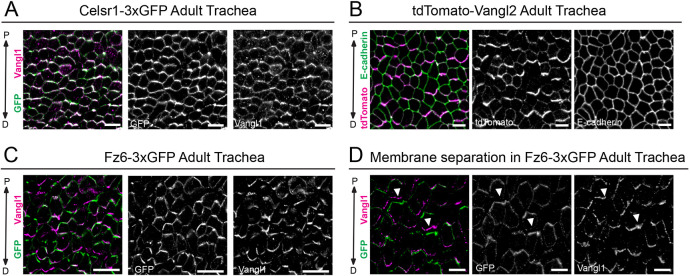
Polarized localization of Celsr1-3xGFP, and mutually exclusive localization of tdTomato-Vangl2 and Fz6-3xGFP to opposite cell edges in the adult trachea. (A-C) Representative planar view of flat-mounted trachea. (A) Homozygous Celsr1-3xGFP adult labeled with antibodies against GFP (green) and Vangl1 (magenta). Note that Celsr1-3xGFP and Vangl1 are asymmetrically localized at proximal-distal (P-D) junctions. Proximal is oriented up. (B) Heterozygous tdTomato-Vangl2 adult labeled with tdTomato (green) and E-cadherin (magenta). tdTomato-Vangl2 is asymmetrically localized along the P-D axis, whereas E-cadherin is uniform around cell edges. (C) Fz6-3xGFP homozygous adult labeled with GFP (green) and Vangl1 (magenta). Fz6-3xGFP and Vangl1 are polarized along the P-D axis. (D) Representative image showing mutually exclusive localization of Fz6-3xGFP (green) and Vangl1 (magenta) to opposing sides of P-D junctions in cells where membranes have separated due to methanol fixation (arrowheads indicate areas showing opposing localization across cell junctions). Scale bars: 10 µm (A,C,D); 5 µm (B).

