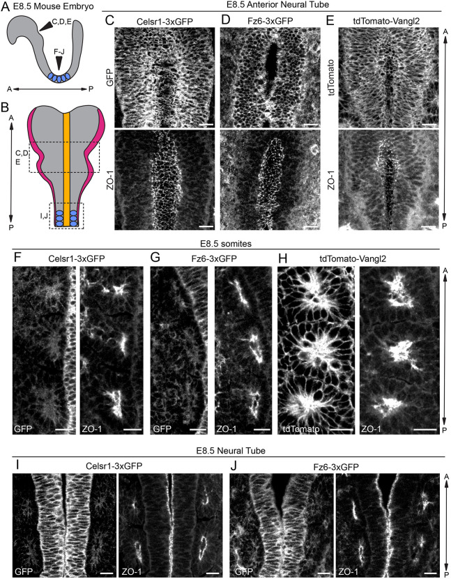
Fig. 7.
Celsr1-3xGFP, Fz6-3xGFP and tdTomato-Vangl2 in the early embryo: neural tube and somites. (A) Schematic of lateral view of E8.5 embryo showing positions of neural tube imaged in C-J and somites (blue). (B) Schematic of dorsal view of E8.5 embryo showing neural folds and somites. Adapted from Brooks et al. (2020). (C-E) Representative images of endogenously-tagged PCP protein localization in the rostral neural tube of E8.5 homozygous embryos labeled with ZO-1 to mark the apical positions of neural epithelial cells. Maximum intensity projections of 5-8 µm are shown. (C) Celsr1-3xGFP (top), ZO-1 (bottom). (D) Fz6-3xGFP (top), ZO-1 (bottom). Note that a single plane was chosen for the Fz6-3xGFP channel to more clearly display its localization. (E) tdTomato-Vangl2 (top), ZO-1 (bottom). (F-H) Representative images showing endogenously-tagged PCP protein localization in the somites of E8.5 homozygous embryos labeled with ZO-1. Three somites from one side of the midline are shown. (F) Celsr1-3xGFP (left) and ZO-1 (right). (G) Fz6-3xGFP (left) and ZO-1 (right). (H) tdTomato-Vangl2 (left) and ZO-1 (right). Note the strong expression of tdTomato-Vangl2 in somites compared with Celsr1-3xGFP and Fz6-3xGFP. (I,J) Representative images of Celsr1-3xGFP and Fz6-3xGFP localization at a more caudal position of the neural tube that has already closed at E8.5. (I) Celsr1-3xGFP (left) and ZO-1 (right). (J) Fz6-3xGFP (left) and ZO-1 (right). Scale bars: 20 µm (C-E); 10 µm (F-J).

