Fig. 1.
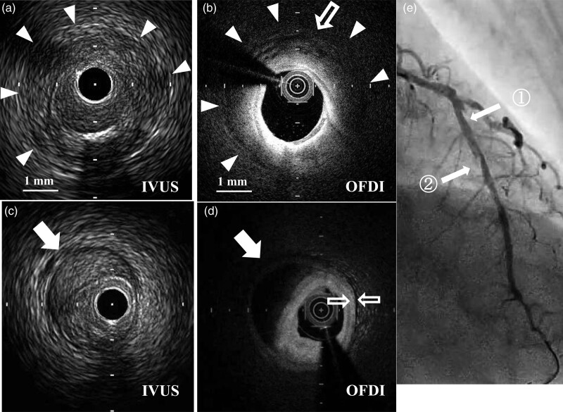
Findings in case 1. (a) After rotational atherectomy, intravascular ultrasound (IVUS) (ViewIT 40 MHz, Terumo, Japan) showed a clearly demarcated low-density circle around the media (triangle arrow), which appears like a ‘halo’ around the vessel. These findings were obtained at the mid-left anterior descending artery just distal to the diagonal branch (E white arrow 1). (b) The optical frequency domain imaging (OFDI) findings of the same site. The arrow head showed clear demarcation of the thickening tunica adventitia. Unlike common hematoma, there was less attenuation of near-infrared (NIR) light and cobblestoning appearance of small low-density aggregates was clearly observed (white blank arrow) at the outermost position of thickened adventitia. (c,d) Common hematoma (white arrow) was observed distally to the lesion (e white arrow 2). IVUS (c) and OFDI (d) images showed very different findings from the images in a and b. First, a common hematoma was clearly demarcated with a low-density band, which may indicate plaque dissection extending into the media (c). Second, due to being rich in pooled blood cells (seen in c), OFDI findings of the hematoma appeared to be a homogenous pitch-black low-density area with strong attenuation of NIR light (d). (e) Angiographic image immediately after rotational atherectomy did not show any signs of coronary perforation (white arrow 1). See video 1, Supplemental digital content 1, http://links.lww.com/MCA/A415.

