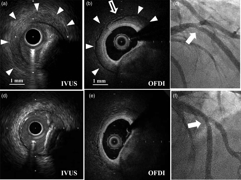
Fig. 2.
Findings in case 2. (a,b) After rotational atherectomy, the same images were obtained in case 2. intravascular ultrasound (IVUS) images (AltaView 40-60 MHz, used set at 50 MHz, Terumo, Japan) and OFDI images (b) closely correlated with case 1. White blank arrow in B indicates a small cobblestoning appearance. (c) Angiographic image immediately after rotational atherectomy did not show any signs of coronary perforation (white arrow). (d) Final IVUS images after the dilatation of drug-coated balloons (DCB 3.5 mm in diameter). The ‘halo’ phenomenon had significantly diminished. (e) OFDI images after the inflation of balloon dilatation (3.0 mm in diameter) before drug-coated ballooning. Unfortunately, final OFDI examination after DCB was not performed, though the thickness of tunica adventitia had normalized at this point, compared with the thickness of adventitia shown in Fig. 1c (white blank arrow). (f) Final angiography. See video 2, Supplemental digital content 2, http://links.lww.com/MCA/A416).

