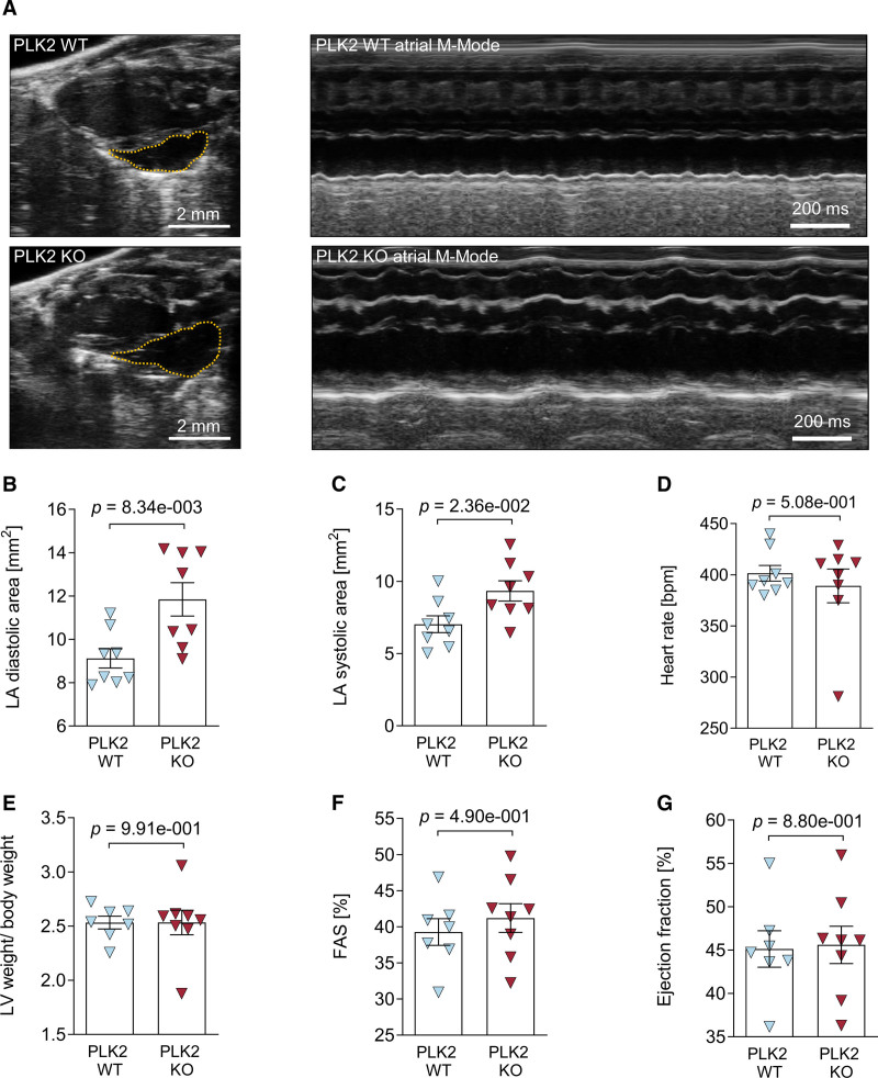
Figure 6.
Echocardiographic characterization of PLK2 (polo-like kinase 2)-deficient mice. Transthoracic echocardiography was performed on isoflurane-anesthetized 8-mo-old PLK2 WT and knockout (KO) mice with a Vevo3100 small animal ultrasound device. A, Representative parasternal long-axis views (left) and M-mode views obtained at the level of the aortic valve (right) recorded from a littermate control (PLK2 WT, upper) and a PLK2-deficient mouse (PLK2 KO, lower); B, Left atrial diastolic area (WT [n=8], KO [n=8]; results are given as mean±SEM, P value was determined by an unpaired, 2-tailed t test). C, Left atrial systolic area (WT [n=8], KO [n=8]; results are given as mean±SEM, P value was determined by an unpaired, 2-tailed t test). D, Heart rate (WT [n=8], KO [n=8]; results are given as mean±SEM, P value was determined by an unpaired, 2-tailed t test). E, Left ventricular weight to body weight ratio (WT [n=7], KO [n=8]; results are given as mean±SEM, P value was determined by a Mann-Whitney U test). F, Fractional area shortening (WT [n=7], KO [n=8]; results are given as mean±SEM, P value was determined by an unpaired, 2-tailed t test). G, Ejection fraction (WT [n=7], KO [n=7]; results are given as mean±SEM, P value was determined by an unpaired, 2-tailed t test).

