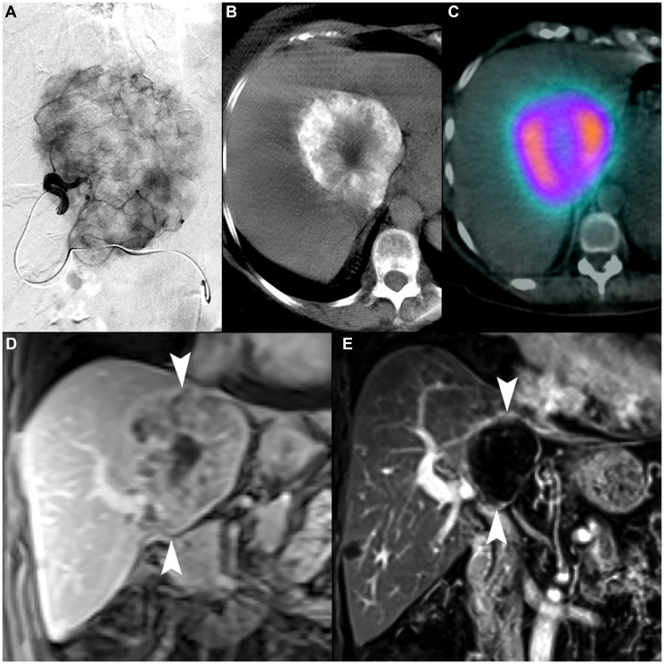
Figure 5. 70-year-old female with intrahepatic cholangiocarcinoma.
(A) Selective arteriography of a 9.2 cm iCCA demonstrating favorable macrovascular conduit for radioembolization. (B) Cone-beam computed tomography showed complete tumor coverage within two angiosomes (second not shown). (C) 99mTc-MAA SPECT/CT demonstrated highly conformal tracer distribution with minimal exposure to non-tumoral liver. The MAA uptake within tumor (including the lower-activity necrotic center) demonstrates an example of favorable microvascular conduit due to overall relative uptake to normal liver parenchyma. (D) Coronal contrast-enhanced MRI image demonstrates the tumor at initial presentation (arrowheads). (E) Coronal contrast-enhanced MRI 3 years after single-session radioembolization with a dose of 476.8 Gy shows a persistent mRECIST complete response and contraction to 4.2 cm (arrowheads).

