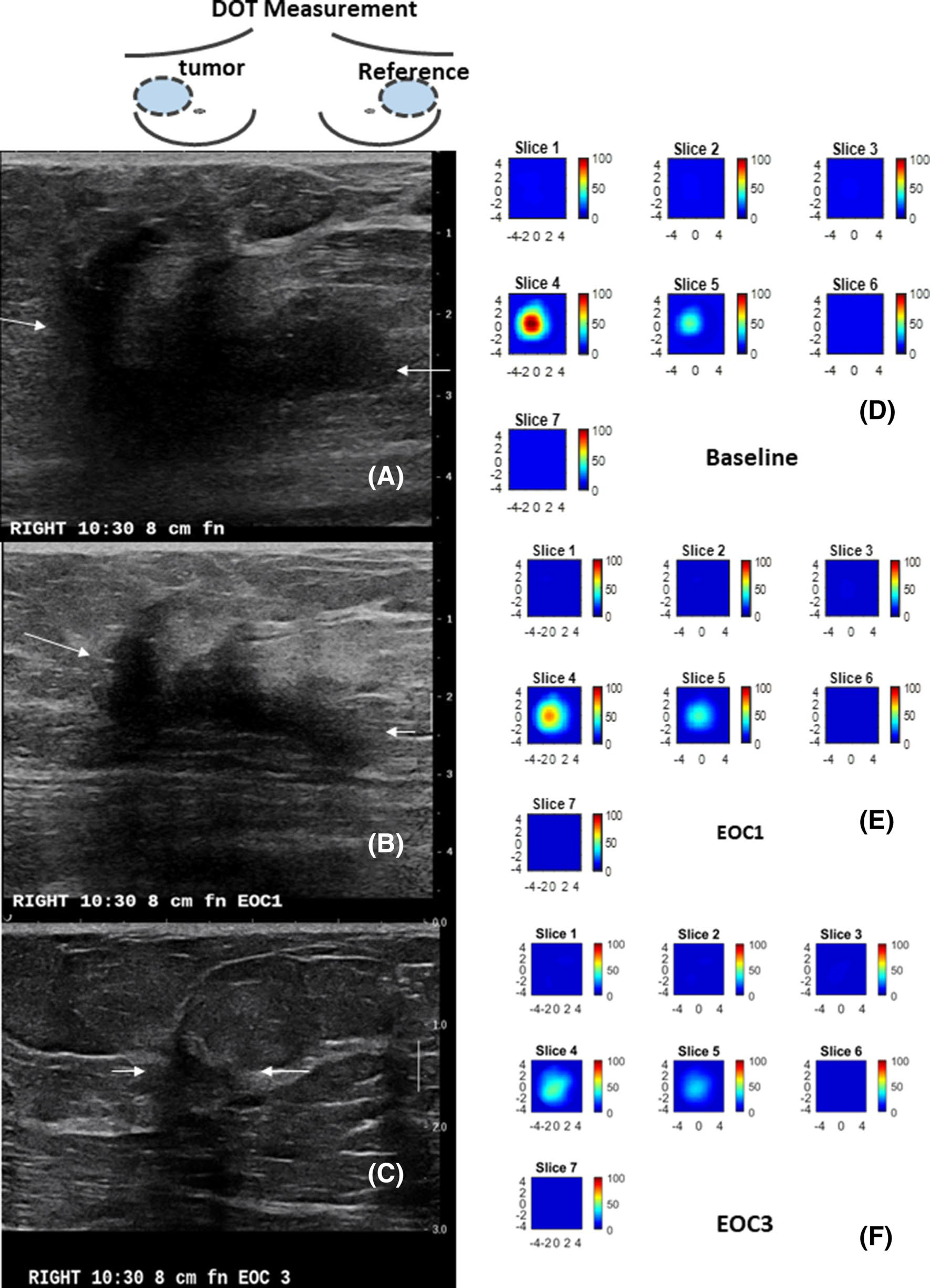
Fig. 4.

A 59 year-old patient with a T3 triple negative cancer and treated with ACT. The US/DOT imaging were performed at baseline, end of cycle 1 (EOC1), 2, 3, 5 and before surgery. a–c are co-registered US images obtained at baseline, EOC1 and EOC3. The largest lesion diameters measured by US were 4.6 cm, 3.4 cm, 1.0 cm. The corresponding %US at EOC1 and EOC 3 were 73.9% and 21.7%. e–f are corresponding HbT maps. Each map has 7 slices reconstructed at depths from 0.5 cm to 3.5 cm with 0.5 cm spacing. Each slice has spatial dimensions of 9 cm by 9 cm. The maximum HbT measured at baseline, EOC1, and EOC3 were 108.7 μM, 73.5 μM, and 45.0 μM. The %HbT were 67.6% and 41.4% at EOC1 and EOC3. The patient achieved pCR with Miller-Payne of 5
