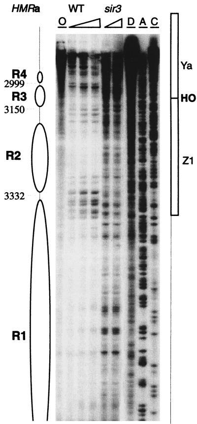
FIG. 2.
HMRa chromatin near the I silencer. Chromatin structure was mapped by primer extension analysis of micrococcal nuclease cleavage sites with primer q35. Wild-type (WT) and sir3 mutant strains are indicated. Triangles represent increasing concentrations of nuclease. Lane O, nuclease-free control; lane D, protein-free DNA subjected to nuclease cleavage; lanes A and C, sequencing reactions to facilitate the identification of locations in the map. The inferred positions of nucleosomes R1 to R4 are indicated. Four precisely positioned nucleosomes are located adjacent to the I silencer. Their structure in the sir3 mutants was disrupted, evidenced by the widespread nuclease accessibility.

