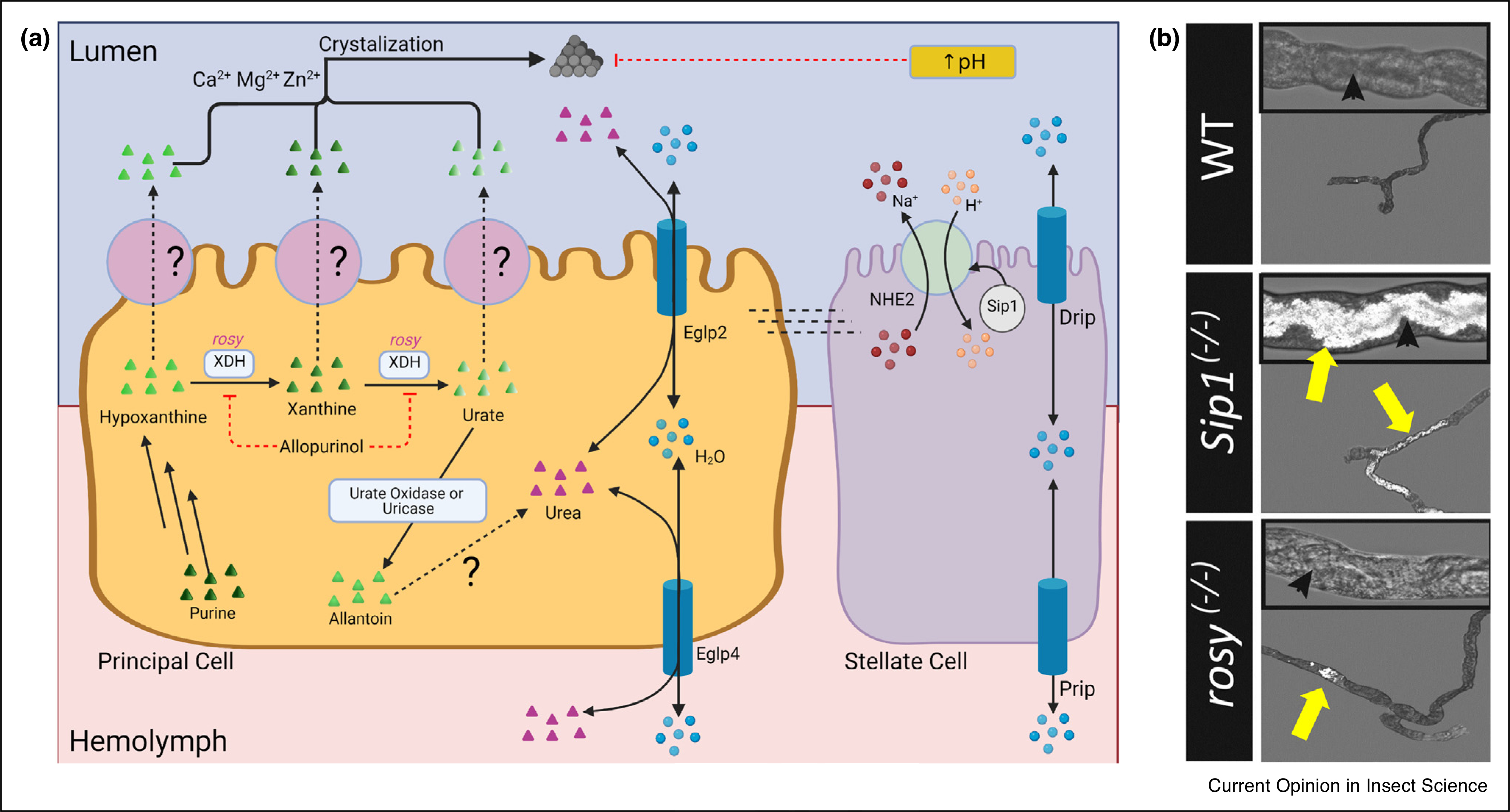
Figure 2. Xanthine and Urate Crystallization in Drosophila renal tubules.

(a) Model of xanthine and urate crystal formation. Purine degradation leads to the production of hypoxanthine. Xanthinuria dehydrogenase/oxidase (Xdh) converts hypoxanthine to xanthine, and then to urate. Urate is then oxidized by urate oxidase (i.e. uricase) to allantoin. Hypoxanthine, xanthine, urate, and allantoin are transported into the lumen by unknown transporter(s) on the apical membrane of principal cells. Allantoin is converted to urea in A. aegypti but this pathway has not yet been identified in other insects. Crystals can form from hypoxanthine, xanthine, or urate with the presence of Zn2+, Ca2+ or Mg2+. Xdh is inhibited by Allopurinol. Crystal formation is inhibited by increased pH from NHE2 and/or Sip-1. Water transport by Prip, Drip, Eglp2, and Eglp4 may influence solute saturation and precipitation. (b) Urate tubuloliths in anterior MT of Sip1(−/−) mutants and xanthine tubuloliths in rosy(−/−) mutants exhibit birefringence due to polarized light (reproduced with permission from Ref. [42•]). Yellow arrows indicate the tubuloliths; black arrowheads mark MT lumen.
