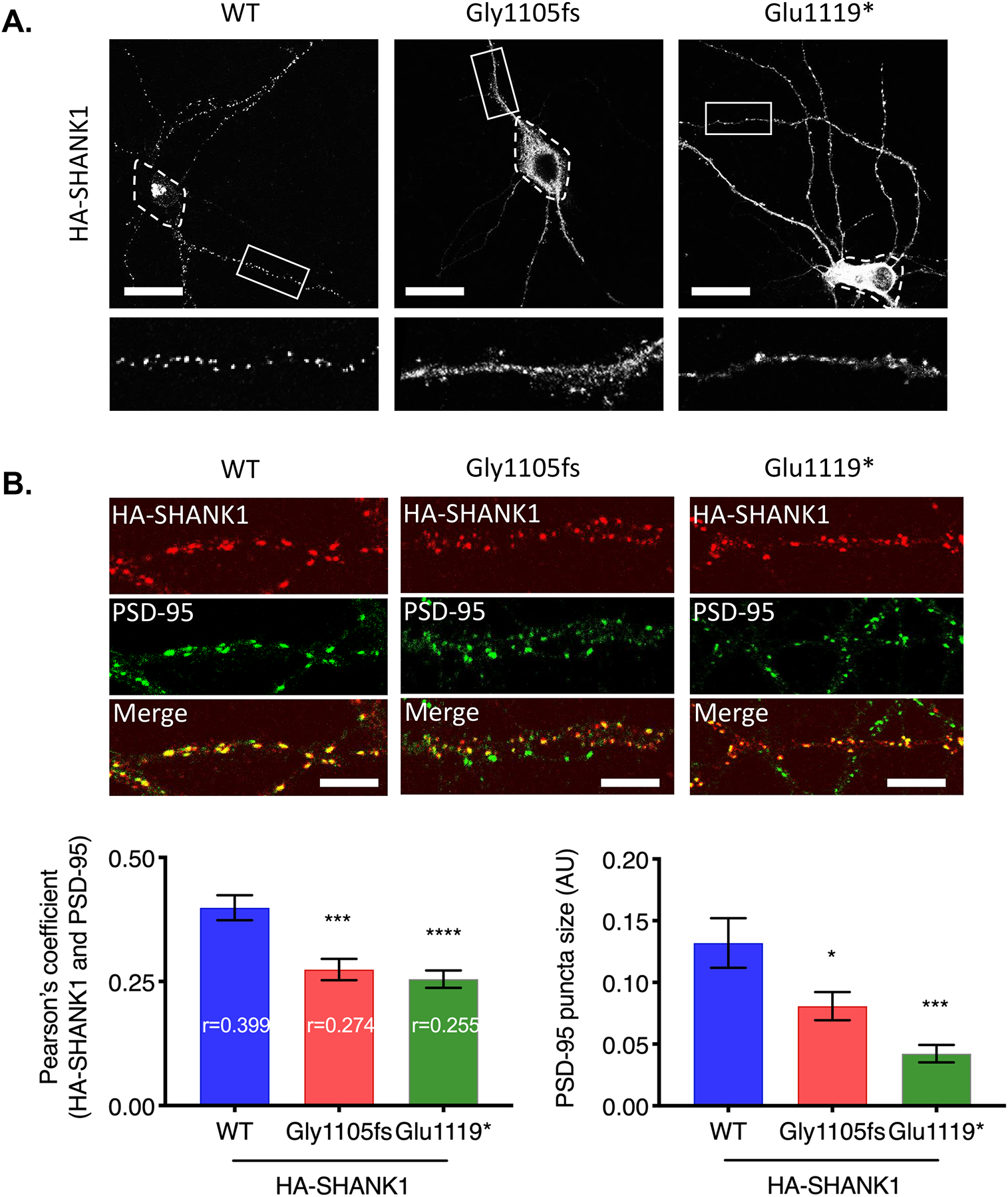
Figure 4. Truncated SHANK1 proteins disrupted synaptic localization.

(A) HA-SHANK1 (WT, Gly1105fs, or Glu1119*) was transfected in cultured rat hippocampal neurons. HA-SHANK1 was labeled with anti-HA and Alexa 555-conjugated secondary antibody (white). Enlarged images of the boxed regions are shown below each panel. Dashed regions indicate soma. (Scale bar, 25 μm.) (B) HA-SHANK1 (WT, Gly1105fs, or Glu1119*) was expressed in cultured rat hippocampal neurons. HA-SHANK1 was labeled with anti-HA and Alexa 555-conjugated secondary antibody (red). Endogenous PSD-95 was labeled with anti-PSD-95 and Alexa 488-conjugated secondary antibody (green). Regions from three dendrites per each neuron were analyzed for Pearson’s coefficient. Graph indicates mean ± SEM (n = 15~19). *P < 0.05, ***P < 0.0005, ****P < 0.0001 using one-way ANOVA with Dunnett’s multiple comparison test. (Scale bar, 5 μm.)
