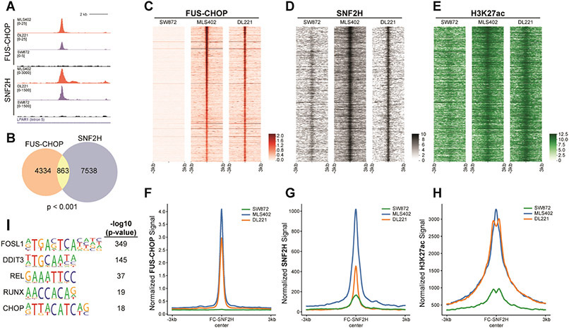
Fig. 3. FUS-CHOP colocalizes with SNF2H at active enhancers.
(A) Representative tracks of a FUS-CHOP SNF2H shared binding site. (B) Venn diagram showing the overlap of FUS-CHOP ROE at enhancers and SNF2H ROE (Permutation test, p < 0.001). (C-E) Heatmaps of FUS-CHOP, SNF2H, and H3K27ac signal densities in human fusion-positive (DL221, MLS402) and fusion-negative (SW872) liposarcoma cell lines. 6-kb windows in each panel are centered on FUS-CHOP SNF2H shared binding sites (n = 863), ranked by significance of overlapping peak calls. (F-H) Average line plots of FUS-CHOP, SNF2H, and H3K27ac signal at ROE bound by both FUS-CHOP and SNF2H. SNF2H signal is increased at these enhancers in the presence of FUS-CHOP. H3K27ac signal is also increased at these enhancers in the presence of FUS-CHOP and SNF2H. The x-axis represents a 6-kb window centered on FUS-CHOP SNF2H binding sites. (I) Enriched motifs found at overlapping FUS-CHOP and SNF2H regions of enrichment. Two biological replicates were used for MLPS cell lines in all experiments.

