
Figure 4. Evaluation of DNA damage defects in NIHCOLE-depleted HCC cells.
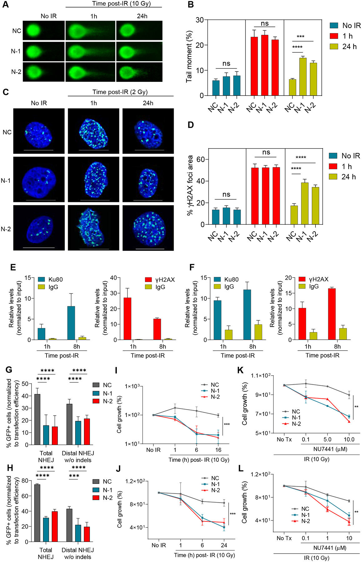
A-D At 24h post-transfection NIHCOLE-depleted, and control cells were either fixed (no IR) or irradiated and fixed at one or 24 hours after irradiation. Cells were then submitted to single-cell electrophoresis for comet scoring. Representative images are shown (A). DNA damage was measured as comet tail-moment (B). Mean ± SEM is shown (n > 90 cells) from two independent experiments. Similarly treated cells were immunostained for γH2AX to detect DNA damage foci (C) Representative images of γH2AX-positive foci (green) and nuclear DNA with DAPI (blue) are shown. Scale bars, 20 μm (D) Percentage of γH2AX foci area over DAPI-stained nuclei. Graph shows mean ± SEM (n > 45 cells) from two independent experiments. E, F RNA immunoprecipitation of NIHCOLE by Ku80 and yH2AX antibodies after radiation. Ku80 and yH2AX antibodies and IgG were used to immunoprecipitate RNA in formaldehyde crosslinked Huh7 (E) and JHH6 (B) cells at early time-points after ionizing irradiation with 2Gy. NIHCOLE was quantified by qRT-PCR. Graph shows mean ± SEM (n = 3 in E, n = 2 in F). G, H Two GFP reporter assays were used to assess NHEJ repair of NIHCOLE-depleted Huh7 (G) and JHH6 (H) cells. The percentage of GFP positive cells was normalized to transfection efficiency. Graph shows mean ± SEM (n = 3 in G, n = 2 in H). I-L Cell growth was measured 48h post-gapmer transfection in control (No IR) or at different times after damage induction by IR in Huh7 (I) and JHH6 (J) cells, or after IR, in mock treated (No Tx) or cells treated with increasing concentrations of DNA-PKcs inhibitor NU7441 in Huh7 (K) and JHH6 (L) cells. Cell number in No IR and No Tx cells was adjusted to 100% to discard the single effect of NIHCOLE depletion on cell growth. Graph shows mean ± SEM (n = 3). The significance of the statistical analysis (one-way ANOVA B, D, G and H; and two-tailed Student’s t test in I-L) is indicated and summarized as: not significant (ns); **<0.01; ***<0.001; ****<0.0001. See also Figure S3.