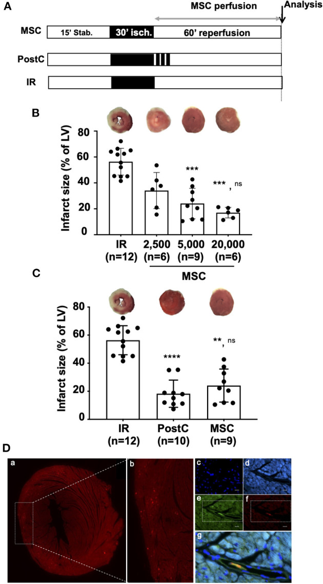
Figure 2.

(A) Isolated hearts perfused ex vivo on the Langendorff system were submitted to perfusion protocol similar to that described in Figure 1A. In the MSC group, reperfusion was achieved with a solution of MSC cells prepared in a Tyrode buffer at various concentrations (2,500; 5,000; or 20,000 cells/mL). For the PostC group, a postconditioning stimulus comprising three cycles of 1 min ischemia-1 min reperfusion was applied at the onset of reperfusion. In the control condition (IR), hearts were reperfused with Tyrode solution alone (control condition). Histological analysis was performed at the end of the protocols for infarct size measurement and immunochemistry. (B) Scatter plots and bars (mean ± SD) were represented for infarct size (in % of LV) in IR (n = 12), MSC 2,500 cells/ml (n = 6), MSC 5,000 cells/ml (n = 9), and MSC 20,000 (n = 6). Representative pictures of TTC-stained LV slices were shown for each group. Statistical analysis was performed using Kruskal-Wallis with the Dunn's post hoc test for multiple comparison. Statistical significance is noted *** for p = 0.0009 (MSC 5,000 vs. IR), *** for p = 0.0003 (MSC 20,000 vs. IR) and ns for p > 0.99 (MSC 20,000 vs. MSC 5,000). (C) Scatter plots and bars (mean ± SD) were represented for infarct size (in % of LV) in IR (n = 12), PostC (n = 10), and MSC (5,000 cells/ml, n = 9). Representative pictures of TTC-stained LV slices were shown for each group. Statistical analysis was performed using Kruskal-Wallis with the Dunn's post hoc test for multiple comparison. Statistical significance is noted **** for p > 0.0001 (PostC vs. IR), ** for p = 0.0018 (MSC vs. IR) and ns for p > 0.99 (PostC vs. MSC). (D) Representative pictures of microscopic observations among 12 (data not shown) for an MSC-treated heart section (a) and (b) corresponding enlarged immunostaining images (Original magnification: x40 oil immersion) showing (c) cell nuclei (DAPI), (d) alpha-actinin, (e) microvessels (isolectin B4), (f) DI-I labeled MSC, and (g) merge allowing to show that MSC are located in the microvessels after 60 min of reperfusion (same time point of infarct size evaluation).
