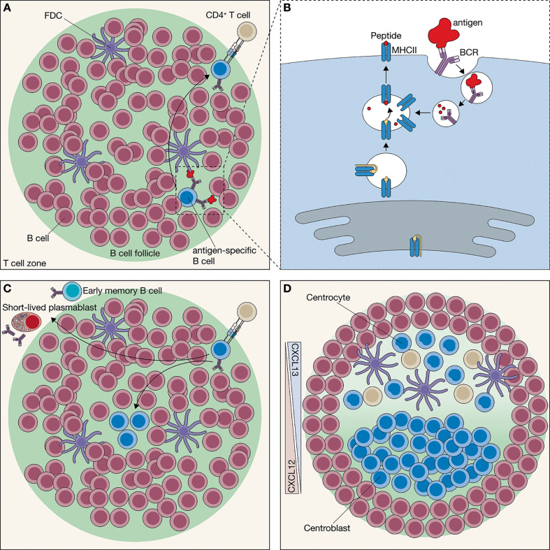
Figure 1.
Initiation of the germinal center (GC) network. (A) In the secondary lymphoid organs, B cells are located in the B cell follicle. Follicular B cells engage with an Antigen (Ag) via their B cell receptor (BCR). Most Ag in the follicle is presented by follicular dendritic cells (FDCs), which retain the Ag for extended periods. Ag engagement partly activates the follicular B cells; however, to become fully activated, it requires interaction with a CD4+ T cells. Ag-engaged follicular B cells localize to the border of the follicle to encounter CD4+ T cells with the same Ag specificity. (B) To interact with the CD4+ T cells, the B cell needs to present Ag-derived peptide fragments through the major histocompatibility complex class II (MHCII). Thereto, Ag engagement results in BCR-mediated endocytosis, followed by Ag degradation and presentation of resulting peptide fragments through MHCII. (C) At the border of the follicle, CD4+ T cells screen many Ag-specific B cells to find the B cell with the same Ag specificity. At the time the CD4+ T cell encounters a follicular B cell with a corresponding Ag specificity, it provides the B cell with help that in turn results in the differentiation towards either short-lived plasmablasts, early memory B cells or GC precursor B cells. GC precursor B cells migrate towards the center of the follicle and starts hyperproliferation. (D) Hyperproliferation drives the formation of the mantel zone, which contains non-activated B cells. As the GC expands the chemokine gradient, mediated by CXCL12 and CXCL13, GC differentiation occurs into two phenotypically distinct zones, the dark zone (DZ) and the light zone (LZ). The CXCL12+ DZ is almost entirely populated by hyperproliferating centroblasts, whereas the CXCL13+ LZ contains FDCs, T follicular helper (Tfh) cells and centrocytes.

