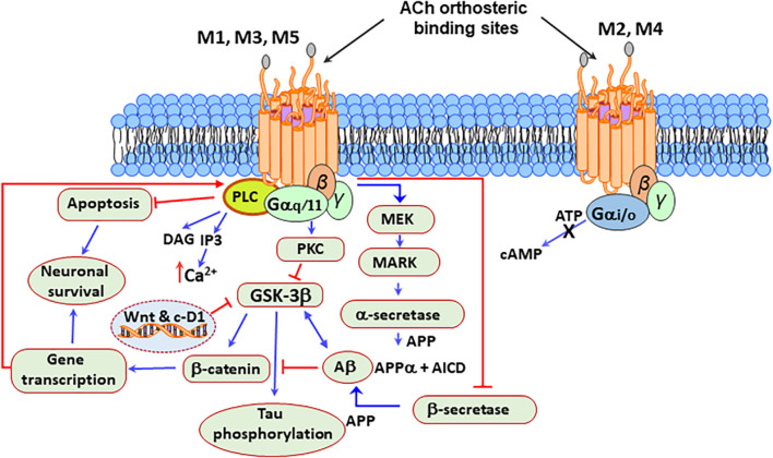
FIGURE 3.
Neuroprotective signaling pathways stimulated by the mAChRs. M1, M3, and M5 mAChRs are associated with the Gq/11 subfamily of G proteins, which are responsible for the increase of cytosolic Ca2+, activation of phospholipase C (PLC) and protein kinase C (PKC), which leads to the production of signaling molecules inositol triphosphate (IP3) and diacylglycerol (DAG). Acetylcholine (ACh) activation of the M2 and M4 receptors, which are associated with the Gi/o subfamily of G proteins, increases the opening time of potassium channels and decreases the production of adenosine-3′,5′-cyclic monophosphate (cAMP). Besides, the stimulation of the M1 mAChRs by agonists or ACh increases the production of sAPPα and decreases the production of amyloid Aβ peptide. Protein kinase C (PKC) and the extracellular signal-regulated protein kinase (ERK)1/2 are involved in this process by activating α-secretases. The activation of the M1 mAChRs counteracts Aβ-induced neurotoxicity via the Wnt signaling pathway, as Aβ inhibits this pathway through the destabilization of β-catenin. In contrast, stimulation of M1 mAChR inactivates glycogen synthase kinase 3 (GSK3β) via PKC activation, thus stabilizing β-catenin and inducing the expression of the Wnt-targeting and cyclin-D1 genes for neuronal survival.

