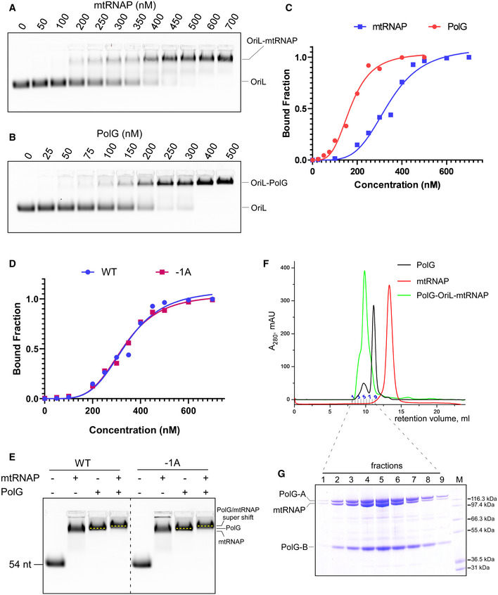
Figure EV3. PolG and mtRNAP assemble into a ternary complex on WT and −1A OriL.

-
A, BEMSA using OriL and mtRNAP (A) and PolG (B).
-
CRelative affinity of mtRNAP and PolG to OriL as observed in A and B.
-
DRelative affinity of mtRNAP to WT and −1A OriL assayed using EMSA.
-
EThe primosome forms on both WT and −1A OriL. EMSA was performed using WT (left) or −1A OriL (right). Note the change of mobility of the labeled species in the presence of both mtRNAP and PolG (“super shift”).
-
FElution profile of PolG‐OriL‐mtRNAP, PolG and mtRNAP during size‐exclusion chromatography on Superdex 200 column.
-
GCoomassie stained SDS–PAGE showing the composition of fractions obtained in size‐exclusion experiment with the primosome complex. Molecular weight markers Mark12 (Invitrogen) are show in lane M.
