Fig. 5.
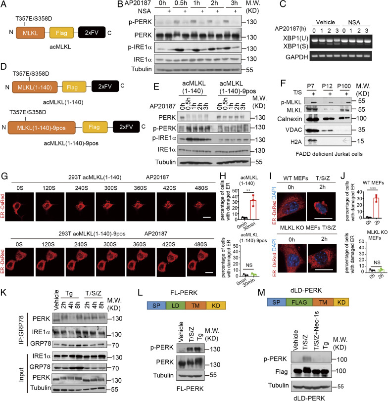
Oligomerized MLKL can activate UPR sensors without disrupting their binding with GRP78. (A) A schematic diagram of the oligomerizable MLKL (acMLKL). T357E/S358D MLKL mutations mimic the phosphorylation by RIPK3. (B and C) acMLKL HT-29 cells were treated with AP20187 (100 nM) and NSA (2.5 μM) as indicated. The cell lysate was collected and analyzed by immunoblotting (B). The cell lysate was collected and analyzed by RT-PCR (C). (D) Schematic representation of the oligomerizable N-terminal part of MLKL (acMLKL-140) and 9 positively charged amino acid MLKL mutants (acMLKL-140–9pos). (E) acMLKL-140 HT-29 cells and acMLKL-140–9pos HT-29 cells were treated with AP20187 (100 nM). The cell lysate was collected and analyzed by immunoblotting. (F) FADD-deficient Jurkat cells were treated with human TNF (50 ng/mL) and SM-164 (100 nM), and the sequential fractions were analyzed by immunoblotting. (G) 293T cells were transfected with the expression constructs of acMLKL (1 to 140), acMLKL (1 to 140)-9pos and ER-DsRed. The cells were treated with AP20187 (100 nM) and ER morphology was detected by time-lapsed imaging. (Scale bar, 10 μm.) (H) Quantification of the percentage of cells with ER puncta in G (>100 cells per group from three independent experiments were assessed). (I) WT MEFs and MLKL KO cells stably expressed with ER-DsRed were treated with mouse TNF (10 ng/mL), SM-164 (100 nM), and z-VAD.fmk (25 μM). The ER morphology was detected by confocal microscopy. (Scale bar, 10 μm.) (J) Quantification of the percentage of cells with ER puncta in I (>100 cells per group from three independent experiments were assessed). (K) HT-29 cells were treated with thapsigargin (500 nM) and human TNF (50 ng/mL), SM-164 (100 nM), and z-VAD.fmk (25 μM) as indicated. The level of IRE1α and PERK binding with GRP78 was detected by coimmunoprecipitation. (L) A schematic diagram of the full-length PERK (FL-PERK) construct. PERK KO HT-29 cells reconstituted with FL-PERK were treated with thapsigargin (500 nM) and human TNF (50 ng/mL), SM-164 (100 nM), z-VAD.fmk (25 μM), and the cell lysate was collected and analyzed by immunoblotting. (M) A schematic diagram of the luminal domain defect PERK (dLD-PERK) construct. PERK KO HT-29 cells reconstituted with dLD-PERK were treated with thapsigargin (500 nM) and human TNF (50 ng/mL), SM-164 (100 nM), z-VAD.fmk (25 μM), and Nec-1s (10 μM). The cell lysate was collected and analyzed by immunoblotting. All data are shown as mean ± SEM; **P < 0.01; ***P < 0.001; NS, not significant; based on two-tailed unpaired Student’s t test (H and J).

