Fig. 1.
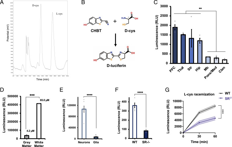
Identification of endogenous d-cysteine in the mammalian brain. (A) Representative chromatogram from HPLC analysis of adult mouse brain extract. (B) Reaction scheme for d-cysteine (d-cys) luciferase assay. Condensation of endogenous d-cys with exogenously added CHBT yields d-luciferin, which is oxidized by d-luciferin–specific luciferase from the firefly P. pyralis to produce light. (C) Luciferase assay measurements of relative d-cys levels in lysates of various regions of adult mouse brain (n = 3). PFC, prefrontal cortex; Thal, thalamus; Str, striatum; Hip, hippocampus; Mb, midbrain; Pons/Med, pons/medulla; and Cbm, cerebellum. (d) Luciferase assay of d-cys levels in postmortem samples of human gray and white matter (n = 2). (E) Luciferase assay of d-cys in primary cortical neuronal and mixed glial cultures (n = 6). (F) Luciferase assay of d-cys in brain lysates from adult wild-type (WT) and serine racemase knockout (SR−/−) mice (n = 5). (G) l-cysteine racemization assay in brain lysates from adult wild-type and SR−/− mice (n = 4). Data are graphed as mean ± SEM **P < 0.01, ***P < 0.001, ****P < 0.0001, ANOVA with post hoc Tukey’s test (C), two-tailed unpaired Student’s t test (D–F), or ANOVA with post hoc Sidak’s test (G).

