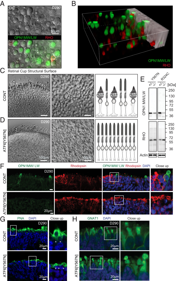
Fig. 1.
ATF6 ACHM patient retinal organoids lack cone structures and cone opsins. (A) Top-down view of DIC microscopy Z-stack series of RHO and cone opsin OPN1MW/LW-labeled retinal organoids shows large round structures on the surface of retinal organoids from normal vision patient control iPSCs (carrying wild-type ATF6). Superimposed immunofluorescence microscopy shows cone opsin OPN1MW/LW-labeling (green) colocalizing with round structures and exclusion of RHO labeling (red). (B) Snapshot from three-dimensional (3D) reconstruction video (Video S1) of confocal fluorescent microscopy images show that round structures on retinal organoid surfaces adopt ovoid morphologies in 3D encapsulating OPN1MW/LW (green) and excluding RHO (red). (C) Images were taken at the same optical magnification; digital zoom was used to demonstrate cellular details of the retinal organoid surface. Overview (Left) and enlargement for detailed view (Middle) DIC microscopy images show abundant cone structures on surface of normal vision patient retinal organoids (CONT). (D) ATF6[Y567N] patient retinal organoids (carrying biallelic inactivating ATF6 alleles) show absence of cone structures with retention of rod structures. Right cartoons in C and D summarize absence of cone structures in ATF6[Y567N] patient retinal organoids. (E) Protein lysates from ATF6[Y567N] and ATF6[R324C] ACHM and control patient retinal organoids (homozygous or heterozygous for ATF6 disease alleles as indicated) were immunoblotted for OPN1MW/LW or RHO. Actin was immunoblotted as a loading control. Protein lysates were combined from five to eight independent retinal organoids of each genotype, and representative immunoblots from three experimental replicates are shown. (F) Confocal fluorescence microscopy images show absence of OPN1MW/LW (green) and preservation of RHO (red) labeling in 290-d (D290)-old ATF6[Y567N] patient retinal organoids (Bottom Rows) compared to normal vision unaffected family member retinal organoid (CONT, Top Rows). DAPI (blue) identifies nuclei. (G) Confocal images show reduced expression of PNA (green) in ATF6[Y567N] (Bottom) retinal organoid compared to CONT (Top) patient retinal organoid. DAPI (blue) labels nuclei. (H) Confocal images show expression of rod transducin/GNAT1 (green) in both control normal vision (CONT, Top) and ATF6[Y567N] patient (Bottom) retinal organoids. For confocal microscopy studies, three independent retinal organoids were analyzed, and representative images are shown. Rhodopsin (RHO), green/red cone opsin (OPN1MW/LW), control (CONT).

