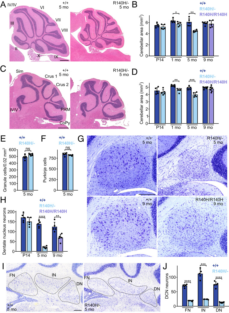
Fig. 2.
Clp1 mutant mice have cerebellar defects. (A and B) H&E-stained midline sagittal sections of cerebellar vermis (A) and mean section area (B). (Scale bar, 500 µm.) (C and D) H&E-stained parasagittal sections of cerebellar hemispheres (C) and mean section area (D). (Scale bar, 500 µm.) (E) Mean granule cell density in lobule IX of vermis. (F) Mean Purkinje cells per midline sagittal section. (G) Dentate nuclei from sagittal sections stained with Cresyl violet. (Scale bar, 200 µm.) (H) Mean large (>100 µm2) dentate nucleus neurons per sagittal section. (I) DCN from coronal sections stained with Cresyl violet. (Scale bar, 200 µm.) (J) Numbers of large (>100 µm2) neurons per coronal hemisection in each nucleus of the DCN. Mean + SD. t tests with Welch’s correction. CoPy, copula pyramidis; DN, dentate nucleus; FN, fastigial nucleus; IN, interposed nucleus; PRM, paramedian lobule; Sim, simple lobule; *P ≤ 0.05; **P ≤ 0.01; ***P ≤ 0.001; ****P ≤ 0.0001; ns, not significant.

