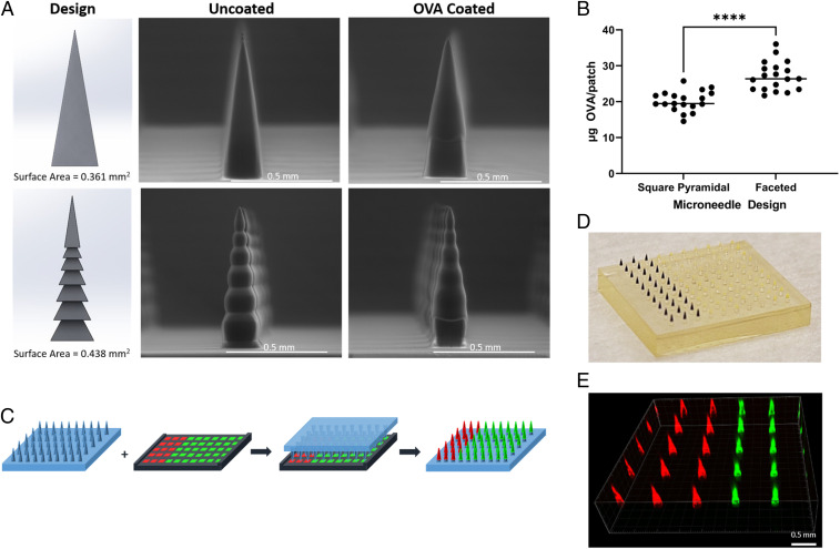
Fig. 1.
CLIP-printed MNs for vaccine formulation. (A) Design and environmental scanning electron microscope (ESEM) images. (Top) Square pyramidal MN. (Bottom) Faceted MN. (B) Ovalbumin coating (n = 19). Data are presented as mean ± SD of individual samples, statistical analysis by unpaired Student’s t tests. ****P < 0.0001. (C) Cargo co-coating scheme. A matching coating mask was used to simultaneously load two cargos onto two sections of needle array. (C) Ovalbumin coating (n = 19). Data are presented as mean ± SD of individual samples, statistical analysis by unpaired Student’s t tests. ****P < 0.0001. (D) Photograph of an OVA–Texas Red and CpG-FITC co-coated MN patch. (E) Fluorescence image of the MN patch in D.

