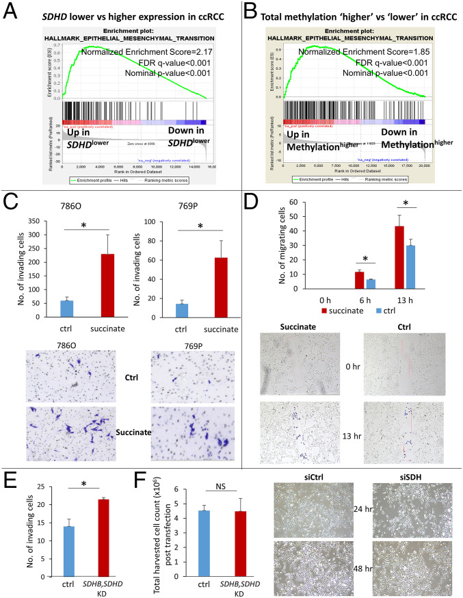
Fig. 5.
Down-regulation of SDH contributes to enhanced invasiveness in ccRCC. (A) Analysis of the KIRC TCGA with the GSEA revealed that lower SDHD is associated with marked enrichment of the Epithelial Mesenchymal Transition Pathway, known to aid invasion and metastasis (NES: 2.17; FDR q value: <0.001). EMT is the most positively enriched pathway in ccRCC tumors with lower SDHD expression. (B) Similarly, higher total methylation in ccRCC tumors is associated with marked enrichment of the EMT pathway (NES: 1.85; FDR q value: <0.001, KIRC TCGA). (C) Succinate treatment (50 μM) of ccRCC cell lines 786-O and 769P resulted in a significant increase in the invasiveness of ccRCC cells as determined by the Matrigel invasion assay (72-h time point, n = 3 for each cell line, data representing mean ± SEM, *P < 0.05). Representative pictures are shown. (D) The Scratch test revealed a significant increase in the migration of ccRCC cells with succinate (50 μM) treatment (n = 2, data representing mean ± SEM, *P < 0.05). Representative pictures shown for 0-, 6-, and 13-h time points. A rectangular area within the scratch (pink mark) was predefined for both succinate and control groups. During analysis, each cell that had invaded the rectangular area was highlighted with a blue dot using the ImageJ imaging software and counted digitally. (E) Knockdown of SDH subunits (SDHB, SDHD) in ccRCC cells (769P) resulted in increased invasiveness (72-h time point, n = 2, data representing mean ± SEM, *P < 0.05; fold change for knockdown of SDHB and SDHD shown in Fig. 4G). There was no difference in harvested cell counts between control and SDH knockdown after transfection in the ccRCC cells. (Please also see SI Appendix, Fig. S23. Knockdown of SDH in ccRCC cells 786-O also resulted in increased invasiveness.)

