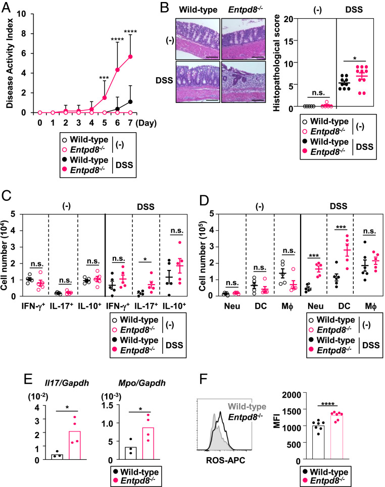
Fig. 2.
Entpd8−/− mice suffer from severe DSS-induced colitis. Mice were orally administered 3% DSS or left untreated for 7 d. (A) The DAI scores of DSS-administered or untreated wild-type (n = 11 or 5, respectively) or Entpd8−/− (n = 12 or 5, respectively) mice (mean values ± SEM). ***P < 0.005; ****P < 0.001. (B) Hematoxylin-eosin staining (Left) and histopathological scores (Right) of colons from DSS-administered or untreated wild-type (n = 9 or 5, respectively) or Entpd8−/− (n = 10 or 5, respectively) mice (mean values ± SEM). (Scale Bars, 100 μm.) *P < 0.05; n.s., not significant. (C) Cell numbers of IFN-γ–, IL-17–, or IL-10–producing CD4+ T cells in the large-intestinal lamina propria from DSS-administered or untreated wild-type or Entpd8−/− mice (n = 5 for all groups) (mean values ± SEM). *P < 0.05; n.s., not significant. (D) Cell numbers of the indicated innate myeloid cell types in the colonic lamina propria from DSS-administered or untreated wild-type (n = 6 or 5, respectively) or Entpd8−/− (n = 6 or 5, respectively) mice (mean values ± SEM). ***P < 0.005; n.s., not significant. (E) Expression levels of Il17 and Mpo in the lamina propria cells from the large intestine of DSS-administered wild-type (n = 3) or Entpd8−/− (n = 4) mice (mean values ± SD). *P < 0.05. (F) Baseline levels of intracellular ROS in colonic Gr-1+ CD11+ cells from DSS-administered wild-type (n = 7) or Entpd8−/− (n = 7) mice (mean values ± SD). All data are from at least two independent experiments. ****P < 0.001.

