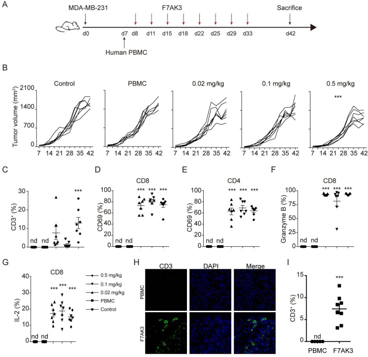
Figure 6.
F7AK3 induces T cell accumulation and reduces tumor burden in a xenogeneic tumor model. (A) Schematic representation depicts the xenograft mouse model. NOD/ShiLtJGpt-Prkdcem26Il2rgem26/Gpt (NCG) immunodeficient mice were subcutaneously injected with 2.5×10⁶ MDA-MB-231 cells. after 7 days, mice were injected with 5×10⁶ human PBMCs intravenously and starting at day 8, mice were treated without or with varying doses of F7AK3 twice a week for four consecutive weeks (n=7 per group). Tumor volumes were measured twice per week. At day 42, mice were sacrificed. (B) Tumor growth curves of all mice were shown, means±SEM. (C) The percentages of CD3+ T cells among single cells of tumor tissues were determined flow cytometry. (D, E) The percentages of CD69+ T cells in CD8+ (D) and CD4+ (E) T cells were assessed by flow cytometry. (F, G) The expression of granzyme B (F) and IL-2 (G) in CD8+ T cells was determined by intracellular staining. (H, I) Immunostaining for CD3 (green) in isolated tumors from PBMCs group and 0.5 mg/kg F7AK3 treated group. Representative images (H) and the percentages of CD3+ T cells among all the tissues cells were quantified manually (I). Data were analyzed by two-way ANOVA analysis with Tukey’s multiple comparisons test (B), one-way ANOVA analysis with Brown-Forsythe test (C–G) and unpaired t-test (H). Mean±SEM; *p<0.05; **p<0.01; ***p<0.001. ANOVA, analysis of variance; IL-2, interleukin 2; Nd, not detected; PBMCs, peripheral blood mononuclear cells.

