. 2021 Aug 17;302(2):319–325. doi: 10.1148/radiol.2021211546

Figure 3:

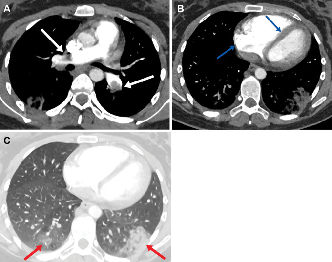
(A) Axial CT pulmonary angiogram image in a 21-year-old woman shows bilateral central pulmonary emboli (arrows) with (B) enlargement of the right heart and flattening of the intraventricular septum in keeping with right heart strain (arrows). (C) Lung window axial CT pulmonary angiogram demonstrates bilateral peripheral areas of opacification in keeping with pulmonary infarcts (arrows).

(A) Axial CT pulmonary angiogram image in a 21-year-old woman shows bilateral central pulmonary emboli (arrows) with (B) enlargement of the right heart and flattening of the intraventricular septum in keeping with right heart strain (arrows). (C) Lung window axial CT pulmonary angiogram demonstrates bilateral peripheral areas of opacification in keeping with pulmonary infarcts (arrows).