. 2021 Aug 17;302(2):319–325. doi: 10.1148/radiol.2021211546

Figure 4:

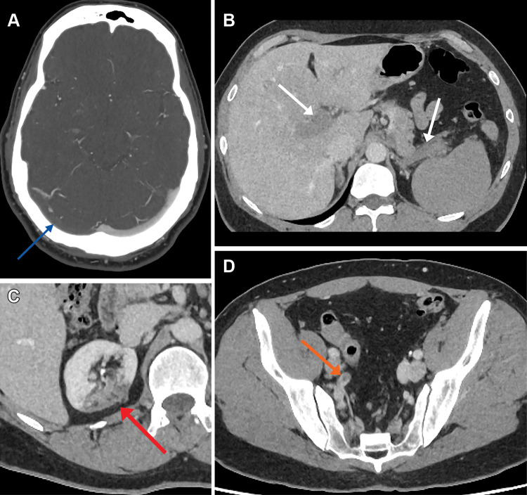
(A) Axial CT venogram image in a 48-year-old man shows a right transverse sinus filling defect in keeping with thrombosis (arrow). (B) Axial portal venous CT of the abdomen and pelvis demonstrates portal vein and splenic vein thromboses (arrows) in addition to (C) right upper pole renal infarct (arrow) and (D) acute right internal iliac artery thrombus (arrow).

(A) Axial CT venogram image in a 48-year-old man shows a right transverse sinus filling defect in keeping with thrombosis (arrow). (B) Axial portal venous CT of the abdomen and pelvis demonstrates portal vein and splenic vein thromboses (arrows) in addition to (C) right upper pole renal infarct (arrow) and (D) acute right internal iliac artery thrombus (arrow).