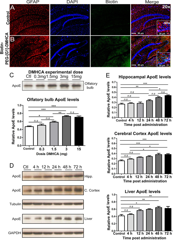
Figure 3.
Biotin-PEG-[G1]-DMHCA intranasally administered reaches the hippocampus after 24 h. In the images, the colocalization of the used markers (red for GFAP/blue for DAPI, nuclei/green for Biotin) can be observed at 20× and 40× (scale bars 50 and 25 μm, respectively) (A,B). Effective intranasal dose analysis in mice: olfactory bulb levels of ApoE evaluated after 24 h by Western Blot (C). Relative levels of ApoE in homogenates of hippocampus, cerebral cortex, and liver; normalizing with values of βIII-tubulin for hippocampus and cerebral cortex, and GAPDH for liver (D,E). Statics analysis was performed using Graph-Pad Prism 6. All probability values were two-tailed; a level of 5% was considered significant. Data are reported as the mean ± SEM.

