Figure 1.
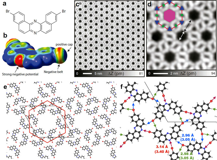
Molecule and self-assembled structure of the multiporous halogen-bonded network grown on Ag(111). (a) Molecular structure of 3,11-dibromo[a, j]phenazine consisting of two sets of bromo-substituted naphthalene moieties fused to a pyrazine core. (b) Electrostatic potential map for the iso-density surface (0.001 e/Bohr3) of the precursor obtained from DFT electrostatic potential calculations, displayed in a red–green–blue colorscale (±15 mV range). (c) Large-scale and (d) close-up view of STM topographies of the network. Three types of pores can be identified within the array that are named with respect to the red hexagon as central (magenta hexagon, area of ∼2.0 nm2), edge (green circles, area of ∼0.18 nm2), and corner (blue triangles, area of ∼0.21 nm2) pores. (e) DFT calculated hexagonal molecular network on Ag(111) marking the red hexagon and the primitive cell. (f) Close-up view indicating the intermolecular distances below 3.5 Å. The numbers in parentheses indicates the sum of the van der Waals radii. Each central pore is defined by six straight C–Br··· N halogen bonds (red arrows), while the edge (corner) pore is defined by two (three) hydrogen bonds [green (blue) arrows]. Measurement parameters: sample voltage V = 10 mV/tunneling current I = 100 pA in (c) and V = 10 mV/I = 10 pA in (d).

