Figure 5.
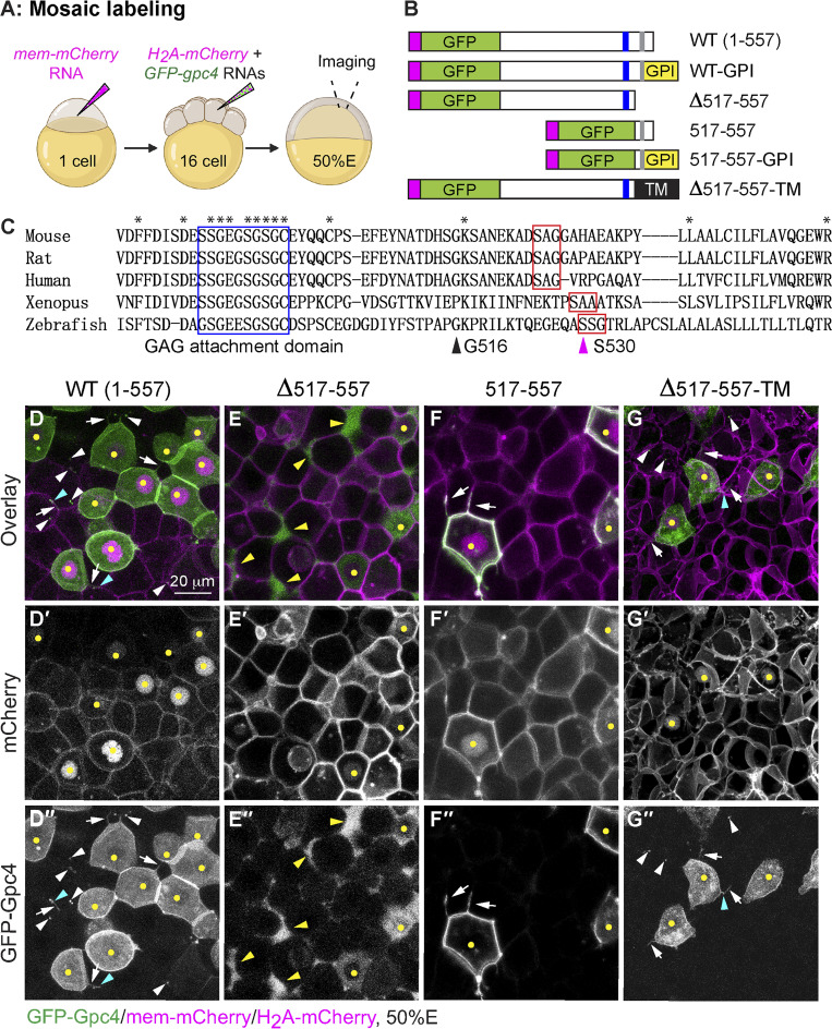
GFP-Gpc4 mutant proteins localize to distinct sites in the zebrafish blastula. (A) Schematic diagram illustrating the mosaic labeling approach for examining the localization of GFP-Gpc4 in vivo. 50%E, 50% epiboly. (B) Schematic diagram of the constructs encoding WT GFP-gpc4 and various mutant forms of the protein. Magenta rectangle, the N-terminal signal peptide; GPI, GPI in the C terminus; blue rectangle, putative GAG attachment domain; gray rectangle, putative GPI attachment site; TM, transmembrane domain of Sdc4. (C) Alignment of C-terminal amino acids of Gpc4 from the indicated species. Asterisks, identical amino acids; blue rectangle, putative GAG attachment domain (G488-C497); red rectangles, putative conserved GPI attachment site and its adjacent residues; magenta arrowhead (at S530), putative GPI attachment site (⍵) in zebrafish; black arrowhead (at G516), the site of fusion to the TM domain. (D–G′′) Confocal images of blastulas, with all cells labeled with mem-mCherry (in magenta, gray in D′–G′) and a subset of cells colabeled with the indicated GFP-Gpc4 constructs (gray in D′′–G′′) and nuclear H2A-mCherry (yellow dots). White arrows, GFP-labeled protrusions; white arrowheads, punctate GFP signal outside of GFP-expressing cells; cyan arrowheads, punctate GFP signal on GFP-labeled protrusions; yellow arrowheads, GFP signal in the extracellular space outside GFP-expressing cells.

