Figure 7.
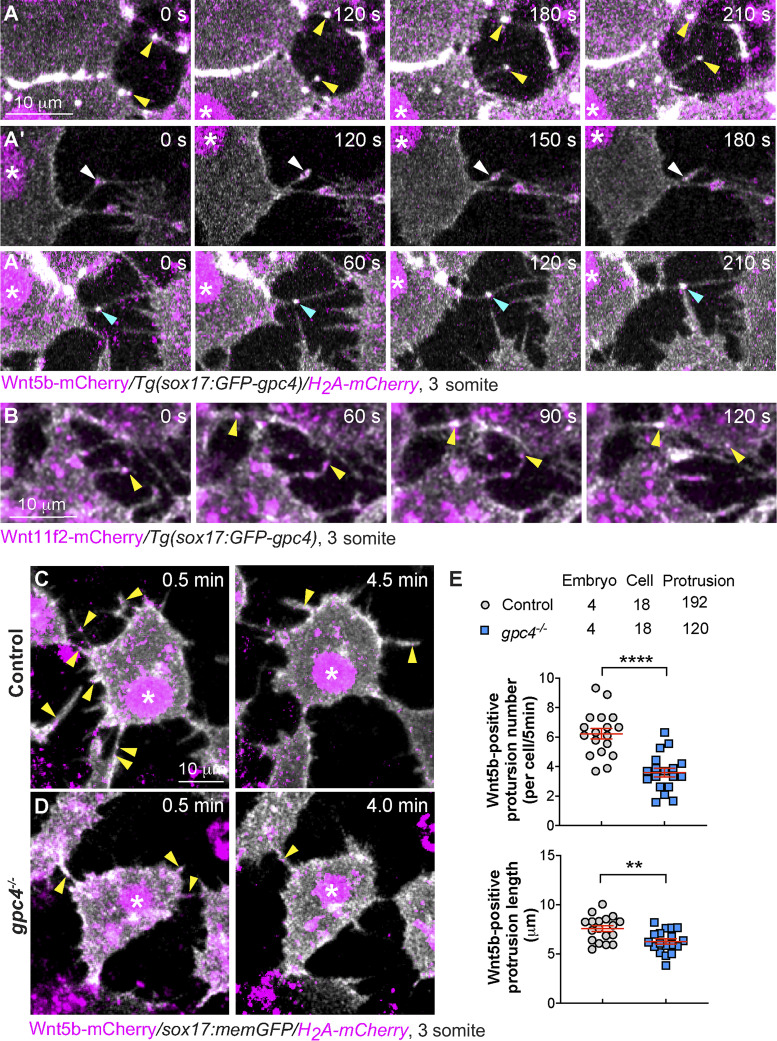
Gpc4 is required for the formation of Wnt-positive filopodia. (A and B) Snapshots from confocal time-lapse images of Tg(sox17:GFP-gpc4/ H2A-mCherry) or Tg(sox17:GFP-gpc4) embryos injected with RNA encoding wnt5b-mCherry (A–A′′, Video 2) or wnt11f2-mCherry (B, Video 3), showing Wnt5b-mCherry or Wnt11f2-mCherry (in magenta) is present on GFP-Gpc4 labeled filopodia (in white). Asterisk, nucleus; yellow arrowheads, Wnt-mCherry at the extending protrusions; white arrowheads, Wnt-mCherry at the retracting protrusions; cyan arrowheads, Wnt-mCherry at protrusions from two cells merging or connected. (C and D) Snapshots from confocal time-lapse images of Tg(sox17:memGFP/H2A-mCherry) embryos injected with wnt5b-mCherry RNA in both control and gpc4−/− embryos (Video 4) showing Wnt5b-mCherry (in magenta, yellow arrowheads) is present on memGFP-labeled filopodia (in white). Asterisk, nucleus. (E) The number and length of protrusions positive for Wnt5b per endodermal cell during a 5-min window, in the indicated embryos, with the number of embryos, cells, and protrusions analyzed indicated. Data are mean ± SEM. **, P < 0.01; ****, P < 0.0001; unpaired Student's t test.

