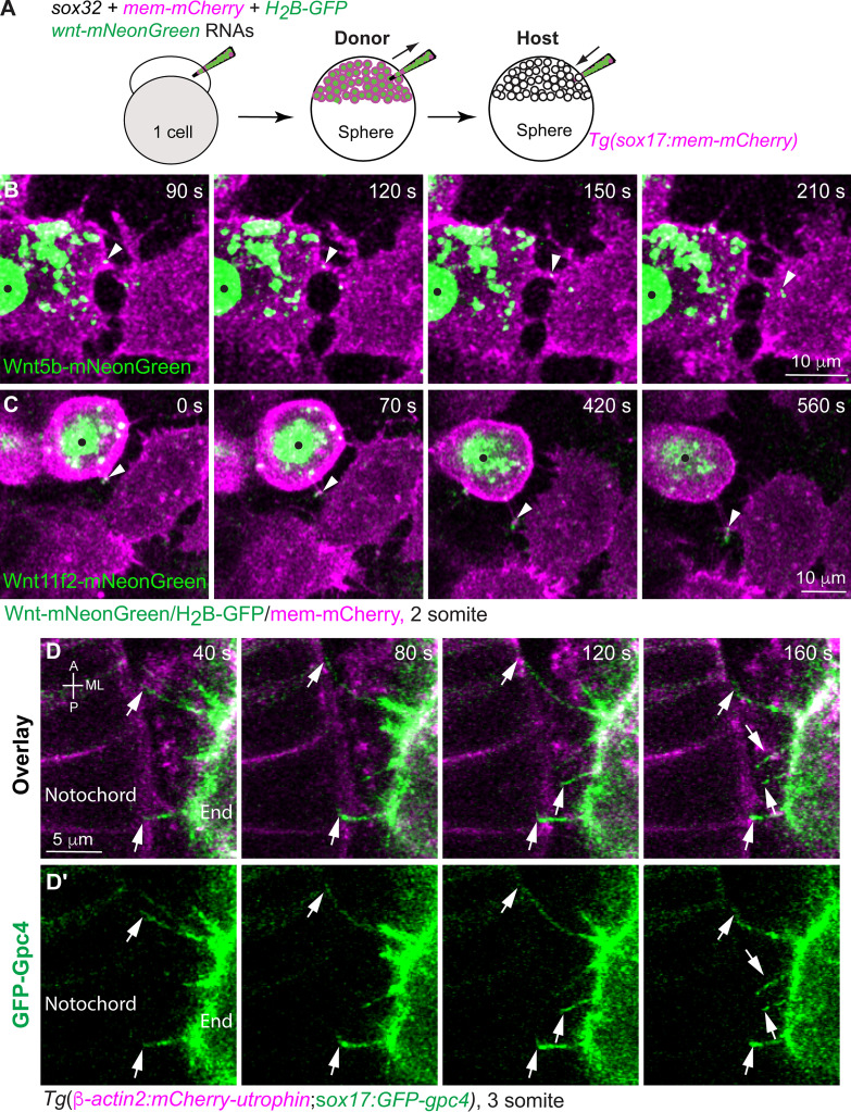
Figure 8.
Endodermal cells extend cellular protrusions to the neighboring endodermal and mesodermal cells. (A) Schematic illustrating endoderm transplantation, in which Wnts-mNeonGreen–expressing donor cells were transplanted into Tg(sox17:mem-mCherry) hosts. (B and C) Snapshots from confocal time-lapse imaging, showing that cellular protrusions extending from wnt5b-mNeonGreen–expressing (B) or wnt11r2-mNeonGreen–expressing donor endodermal cells (C; black dots) transport Wnt5b-mNeonGreen (B) or Wnt11f2-mNeonGreen (C) to neighboring endodermal cells (Video 6). Arrowheads, Wnt puncta on protrusions. (D–D′) Snapshots from confocal time-lapse imaging on Tg(β-actin2:mCherry-utrophin;sox17:GFP-gpc4) embryos in which the plasma membrane of notochord cells and endodermal cells was labeled with mCherry (Video 7). Images were taken on the region where endodermal cells and the notochord were in close proximity, showing that GFP-Gpc4–labeled protrusions from endodermal cells (D′, arrows) extended toward and contacted mCherry-Utrophin–expressing notochord cells (D). End, endoderm cell.

